. 2005 Aug 15;170(4):571-82.
doi: 10.1083/jcb.200501097.

An ECT2-centralspindlin complex regulates the localization and function of RhoA

Affiliations

An ECT2-centralspindlin complex regulates the localization and function of RhoA

Ozlem Yüce et al. J Cell Biol. .

Abstract

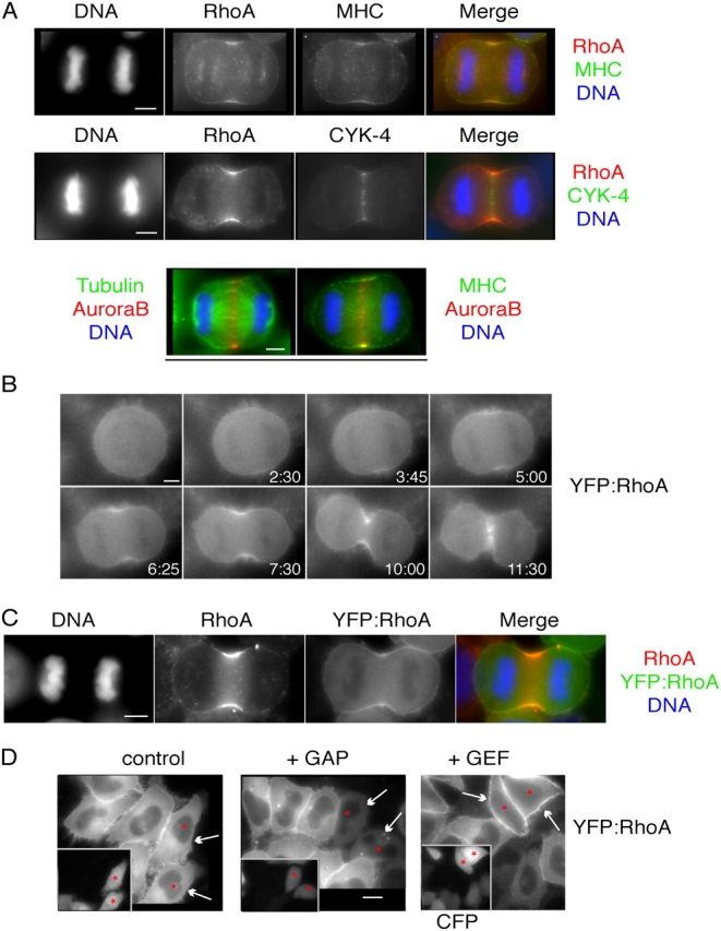
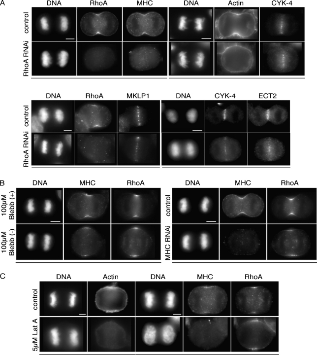
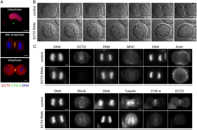
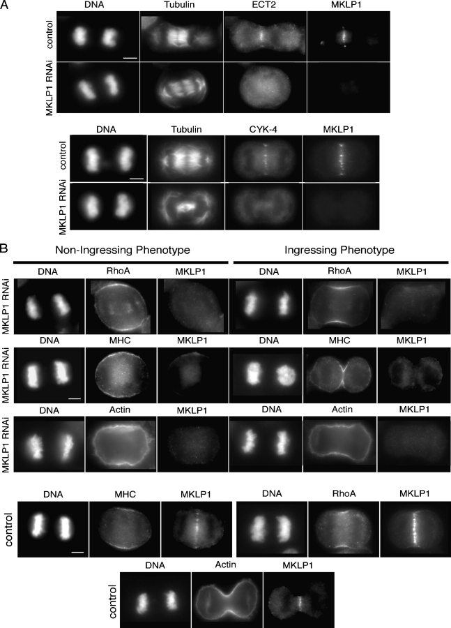
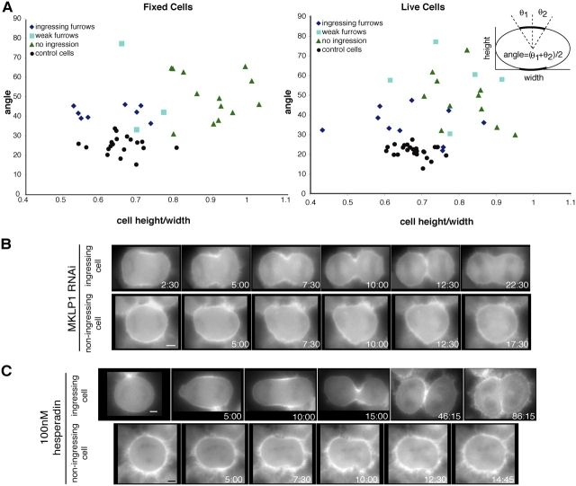
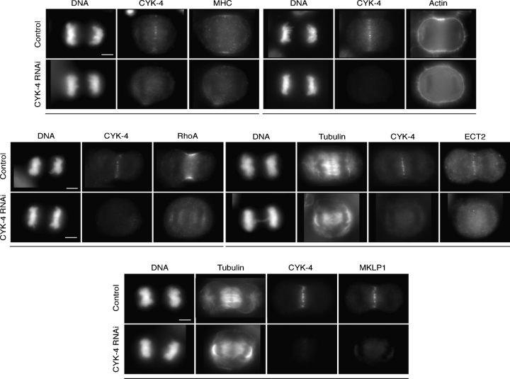
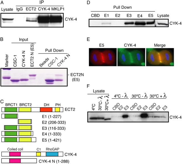
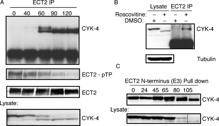
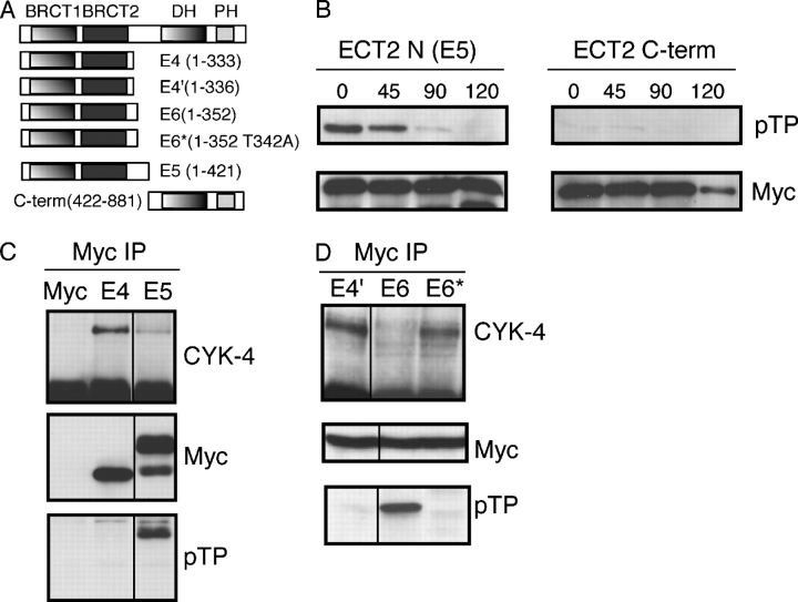
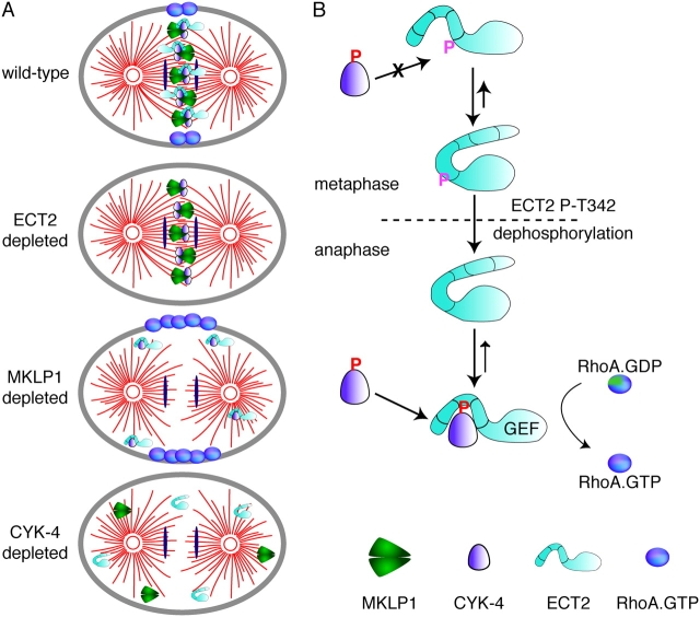
In anaphase, the spindle dictates the site of contractile ring assembly. Assembly and ingression of the contractile ring involves activation of myosin-II and actin polymerization, which are triggered by the GTPase RhoA. In many cells, the central spindle affects division plane positioning via unknown molecular mechanisms. Here, we dissect furrow formation in human cells and show that the RhoGEF ECT2 is required for cortical localization of RhoA and contractile ring assembly. ECT2 concentrates on the central spindle by binding to centralspindlin. Depletion of the centralspindlin component MKLP1 prevents central spindle localization of ECT2; however, RhoA, F-actin, and myosin still accumulate on the equatorial cell cortex. Depletion of the other centralspindlin component, CYK-4/MgcRacGAP, prevents cortical accumulation of RhoA, F-actin, and myosin. CYK-4 and ECT2 interact, and this interaction is cell cycle regulated via ECT2 phosphorylation. Thus, central spindle localization of ECT2 assists division plane positioning and the CYK-4 subunit of centralspindlin acts upstream of RhoA to promote furrow assembly.


Figures

Figure 1.
Figure 1.
RhoA localizes to the equatorial cell cortex in early anaphase. (A) HeLa cells were fixed with TCA and stained with antibodies directed against RhoA (red) and either myosin IIB heavy chain or CYK-4 (green). HeLa cells fixed with methanol were stained with antibodies directed against aurora B (red) and tubulin or myosin II (green). (B) YFP:RhoA concentrates at the equatorial cortex before furrow formation. A HeLa cell stably expressing YFP:RhoA is shown at successive time points during cytokinesis (Video 2). This cell was treated with lamin B siRNA. (C) YFP:RhoA colocalizes with endogenous RhoA. Cells stably expressing YFP:RhoA were fixed with TCA and stained with antibodies directed against RhoA (red) and YFP (green). The anti-RhoA antibody does not cross react with YFP:RhoA. (D) Cortical localization of YFP:RhoA increases upon RhoA activation and decreases upon RhoA inhibition (arrows). The GAP domain of RhoGAP1 or the GEF domain of ECT2 were coexpressed with CFP in YFP:RhoA cells. CFP-positive cells (inset) are indicated (asterisks). Images obtained from the same cell are underlined. Bars: (A–C) 5 μm; (D) 10 μm.
Figure 2.
Figure 2.
RhoA regulates myosin II accumulation and localizes in the absence of myosin II or F-actin. (A) RhoA is required for accumulation of myosin II and actin. HeLa cells depleted of RhoA were fixed and stained with the indicated antibodies and rhodamine phalloidin. (B) Neither myosin activity nor myosin is required for RhoA localization. HeLa cells were treated with 100 μM Blebbistatin (−, active) or Blebbistatin (+, inactive) for 45 min or treated with siRNAs to myosin heavy chain before fixation and immunofluorescence. (C) F-actin is not required for RhoA localization. HeLa cells treated with 5 μM LatA for 45 min were fixed and stained with the indicated antibodies and rhodamine phalloidin. Images obtained from the same cell are underlined. Bars, 5 μm.
Figure 3.
Figure 3.
ECT2 localizes to the central spindle and regulates RhoA localization and cytokinesis. (A) ECT2 localizes to the central spindle and colocalizes with the centralspindlin complex. (B) ECT2 is required for furrow formation. ECT2 depleted or control cells undergoing cytokinesis were imaged by DIC microscopy (Videos 3 and 4). (C) Neither RhoA, myosin, nor actin localize to the cell cortex in ECT2-depleted cells, but the central spindle assembles normally. ECT2-depleted or control cells were fixed and stained with the indicated antibodies or rhodamine phalloidin. Images obtained from the same cell are underlined. Bars: (A, top) 10 μm; (A–C) 5 μm.
Figure 4.
Figure 4.
MKLP1 is required for ECT2 localization. (A) Cells depleted of MKLP1 were fixed and stained with the indicated antibodies. (B) MKLP1 restricts the localization of RhoA, but it does not block cortical localization of RhoA. MKLP-depleted cells were fixed and stained with the indicated antibodies or rhodamine phalloidin. Cells that exhibit the two classes of cytokinesis phenotypes are shown. Bars, 5 μm.
Figure 5.
Figure 5.
Expansion of cortically localized RhoA upon MKLP1 depletion and aurora inhibition. (A) Quantitation of RhoA localization in MKLP1-depleted cells and control cells. Cell morphology and the extent of RhoA localization in fixed and live cells were measured as indicated. Fixed cells were scored for the presence of a furrow and live cells were scored for furrow formation during the course of the recording. Fixed cells with partly condensed, and segregated DNA were measured. Live cells were measured when RhoA localization was most restricted. (B) YFP:RhoA-expressing cells were depleted of MKLP1 by RNAi. YFP:RhoA was imaged and shown at successive time points during cytokinesis. Two phenotypes were observed. Either RhoA remained somewhat restricted and these cells formed furrows that regress; otherwise RhoA spread out along the cell cortex and furrows did not form (Videos 6 and 7). (C) HeLa cells stably expressing YFP:RhoA were treated with 100 nM Hesperadin and YFP:RhoA was imaged during cytokinesis (Videos 8 and 9). Bars, 5 μm.
Figure 6.
Figure 6.
CYK-4 is required for central spindle formation and is essential for active RhoA localization. Cells depleted of CYK-4 were fixed and stained with the indicated antibodies or rhodamine phalloidin. YFP:RhoA localization was also abolished in CYK-4–depleted cells (Video 10). Images obtained from the same cell are underlined. Bars, 5 μm.
Figure 7.
Figure 7.
CYK-4 and ECT2 directly interact and associate in vivo in a phosphorylation-dependent manner. (A) ECT2, CYK-4, and MKLP1 were precipitated and the immunoprecipitates were subjected to Western blotting with anti–CYK-4 antibodies. (B) Recombinant, soluble ECT2 E5 was incubated with CBD beads or CBD beads bound to CBD-CYK-4-N or CBD-CSC-1. Bound proteins were analyzed by Coomassie staining. (C) Schematic showing the boundaries of deletion constructs. (D) The tandem BRCT domains are sufficient for efficient binding of CYK-4. E. coli–expressed derivatives bound to chitin beads were used to pull-down proteins from a mitotic cell lysate. Bound proteins were analyzed by Western blotting with anti–CYK-4 antibodies. (E) The ECT-2 truncation derivative E5 colocalizes with CYK-4. Myc-tagged ECT2 E5 construct was transfected into HeLa cells, fixed, and stained for myc and CYK-4. (F) The association between CYK-4 and the ECT2 BRCT domains is phosphodependent. Mitotic cell lysates were treated with λ phosphatase before binding to chitin beads loaded with the indicated proteins. Bound proteins were analyzed by Western blotting with anti–CYK-4 antibodies.
Figure 8.
Figure 8.
The association between CYK-4 and ECT2 is cell cycle regulated. (A) Time course analysis of the association of CYK-4 and ECT2 during mitotic exit. HeLa cells were arrested in metaphase with nocodazole and released. Lysates were prepared at the indicated times and ECT2 was immunoprecipitated. Immunoprecipitates were Western blotted with anti–CYK-4 and anti–phospho-Thr-Pro antibodies to detect phosphorylation of ECT2. (B) CDK1 activity antagonizes the interaction between CYK-4 and ECT2. Metaphase-arrested HeLa cells were treated with Roscovitine for 30 min, ECT2 was immunoprecipitated, and the immunoprecipitates were Western blotted with anti–CYK-4 antibodies. (C) Recombinant ECT2 pulls down CYK-4 equally efficiently in metaphase and anaphase. HeLa cells were arrested in metaphase with nocodazole and released from the arrest. Lysates were prepared at the indicated times and ECT2 binding proteins were recovered with chitin beads loaded with the E3 fusion protein. Bound proteins analyzed by Western blotting with anti–CYK-4 antibodies.
Figure 9.
Figure 9.
Identification of a phosphorylation site that regulates the CYK-4–ECT-2 interaction. (A) Schematic of the ECT2 constructs used. (B) The NH2- and COOH-terminal halves (E5 and C-term) of ECT2 were transfected into HeLa cells, which were then arrested in metaphase with nocodazole. Cells were released from the block and collected at the indicated times. The myc-tagged ECT2 constructs were immunoprecipitated and Western blotted with anti-myc and anti–phospho-Thr-Pro antibodies. (C) ECT2 fragments are differentially competent to bind CYK-4 during metaphase. The differential association inversely correlates with reactivity with anti–phospho-Thr-Pro antibody. ECT2 constructs were transfected into HeLa cells, which were then arrested in metaphase with nocodazole. Lysates were prepared and the myc-tagged ECT2 constructs were immunoprecipitated and Western blotted with anti-CYK-4, anti-myc, and anti–phospho-Thr-Pro antibodies. (D) Identification of threonine 342, which, when mutated to alanine, prevents TP phosphorylation and allows ECT2 to bind CYK-4 during metaphase. This experiment was performed as described in C.
Figure 10.
Figure 10.
The phenotypes of depleted cells and the regulated association of CYK-4 and ECT2. (A) A schematic model summarizing the phenotypes of cells depleted of MKLP1, CYK-4, and ECT2. (B) A model for the regulated association of CYK-4 and ECT2. See text for details.

References

    1. Adams, R.R., A.A. Tavares, A. Salzberg, H.J. Bellen, and D.M. Glover. 1998. pavarotti encodes a kinesin-like protein required to organize the central spindle and contractile ring for cytokinesis. Genes Dev. 12:1483–1494. - PMC - PubMed
    1. Aghazadeh, B., W.E. Lowry, X.Y. Huang, and M.K. Rosen. 2000. Structural basis for relief of autoinhibition of the Dbl homology domain of proto-oncogene Vav by tyrosine phosphorylation. Cell. 102:625–633. - PubMed
    1. Alberts, A.S. 2001. Identification of a carboxyl-terminal diaphanous-related formin homology protein autoregulatory domain. J. Biol. Chem. 276:2824–2830. - PubMed
    1. Bement, W.M., H.A. Benink, and G. von Dassow. 2005. A microtubule-dependent zone of active RhoA during cleavage plane specification. J. Cell Biol. 170:91–101. - PMC - PubMed
    1. D'Avino, P.P., M.S. Savoian, and D.M. Glover. 2004. Mutations in sticky lead to defective organization of the contractile ring during cytokinesis and are enhanced by Rho and suppressed by Rac. J. Cell Biol. 166:61–71. - PMC - PubMed

Publication types

MeSH terms