Comparative Study
. 2005 Aug 23;102(34):12047-52.
doi: 10.1073/pnas.0505207102. Epub 2005 Aug 15.

X-ray damage to the Mn4Ca complex in single crystals of photosystem II: a case study for metalloprotein crystallography

Affiliations
Comparative Study

X-ray damage to the Mn4Ca complex in single crystals of photosystem II: a case study for metalloprotein crystallography

Junko Yano et al. Proc Natl Acad Sci U S A. .

Abstract

X-ray absorption spectroscopy was used to measure the damage caused by exposure to x-rays to the Mn(4)Ca active site in single crystals of photosystem II as a function of dose and energy of x-rays, temperature, and time. These studies reveal that the conditions used for structure determination by x-ray crystallography cause serious damage specifically to the metal-site structure. The x-ray absorption spectra show that the structure changes from one that is characteristic of a high-valent Mn(4)(III(2),IV(2)) oxo-bridged Mn(4)Ca cluster to that of Mn(II) in aqueous solution. This damage to the metal site occurs at a dose that is more than one order of magnitude lower than the dose that results in loss of diffractivity and is commonly considered safe for protein crystallography. These results establish quantitative x-ray dose parameters that are applicable to redox-active metalloproteins. This case study shows that a careful evaluation of the structural intactness of the active site(s) by spectroscopic techniques can validate structures derived from crystallography and that it can be a valuable complementary method before structure-function correlations of metalloproteins can be made on the basis of high-resolution x-ray crystal structures.


Figures
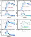
Fig. 1.
Fig. 1.
Mn XANES of PS II versus x-ray dose and XANES of inorganic model compounds. (A) Mn K edge shift of PS II crystals as a function of x-ray dose at 13.3 keV (0.933 Å) and 100 K (Upper) and the difference spectra (Lower). The spectrum at the highest inflection point energy is from an undamaged PS II crystal. The x-ray doses used for exposure are 0.14, 0.21, 0.25, 0.54, 0.95, 2.3, and 5.0 × 10¹⁰ photons per μm2 (light-blue to black lines). An average dose of ≈3.5 × 10¹⁰ photons per μm2 was used for x-ray diffraction studies. The exposure was at 100 K, and all XANES was collected at 10 K at low dose (1 × 10⁷ per μm2). XANES and difference spectra show that the increase in amplitude at ≈6,552 eV provides definite evidence for the photoreduction to Mn(II) in PS II crystals by exposure to x-rays (see C). At a dose of 2.3 × 10¹⁰ photons per μm2, equal to 66% of the average dose used for diffraction measurements, 80% of the Mn in PS II is present as Mn(II). (B) A similar trend in the XANES (Upper) and the difference spectra (Lower) is seen for PS II solutions. The x-ray doses used for exposure were 0.03, 0.05, 0.10, 0.17, 0.32, 0.76, and 1.4 × 10¹⁰ photons per μm2 (light-blue to black lines). At a dose of 1.4 × 10¹⁰ per μm2, equal to 40% of the average dose used for diffraction measurements, 90% of the Mn in PS II is present as Mn(II). (C) The changes in the XANES spectra from an intact PS II sample as a function of addition of 10% increments of the XANES spectrum of aqueous Mn(II) (Upper) and the corresponding difference spectra (Lower) (light-blue to black lines). The similarity between the difference spectra in A, B, and C is striking, showing the photoreduction of the Mn(III2,IV2) in native PS II to Mn(II). (D) A comparison of Mn K edge spectra from two tetranuclear complexes, [Mn4O3(OAc)4(dbm)3] (35, 36) and [(Mn2O2)2(tphpn)2](ClO4)4 (37, 38), in oxidation states Mn4(III3,IV) and Mn4(III2,IV2) similar to those in intact PS II and from Mn(II) in aqueous solution.
Fig. 2.
Fig. 2.
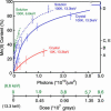
Increasing Mn(II) content in PS II due to radiation damage. (Solid blue line) Mn(II) content in PS II crystals as a function of x-ray irradiation at 13.3 keV (0.933 Å). The irradiation was carried out at 100 K. The conditions are similar to those during x-ray diffraction data collection. The dose on the abscissa is given in grays and in photons per unit area, units that are commonly used for crystallography and spectroscopy experiments, respectively. The points on the curve represent samples that received between 0.14 and 5.0 × 10¹⁰ photons per μm2. At 66% of the dose (2.3 × 10¹⁰ photons per μm2) compared with the representative average dose (3.5 × 10¹⁰ photons per μm2) used for crystallography, PS II crystals contain ≈80% Mn(II). (Dashed blue line) The damage profile for PS II solution samples is very similar to that seen for crystals, although it is slightly higher for the same dose. EXAFS spectra of samples shown in Fig. 3 are at levels of damage denoted on the right by A, B, C, D, and E. (Dashed green line) The generation of Mn(II) is considerably greater when the x-ray irradiation is at 6.6 keV (1.89 Å), which is the energy at which the anomalous diffraction measurements for PS II were conducted. (Solid blue line) The Mn(II) produced by damage in crystals is considerably decreased when the irradiation is conducted at 10 K, providing a method that could be used to mitigate the effects of radiation damage during crystallography measurements.
Fig. 3.
Fig. 3.
Spectral changes of PS II Mn EXAFS due to radiation damage with FTs (Left) and the k3-space EXAFS (Right). (Left) The FT of the EXAFS spectrum from an intact PS II solution sample is on top (red). The three Fourier peaks are characteristic of a bridged Mn4Ca complex, with peak I from bridging Mn-oxo and Mn-terminal ligand atoms, peak II from Mn–Mn distances at 2.7 Å characteristic of di-μ-oxo-bridged moieties, and peak III from mono-μ-oxo-bridged Mn–Mn distances at 3.3 Å and Mn–Ca distances at 3.4 Å. The FTs from PS II solution samples exposed to 0.05, 0.1, 0.3, and 1.4 × 10¹⁰ photons per μm2 at 13.3 keV and 100 K and containing 25% (blue), 35% (dark blue), 45% (green), and 90% (black) photoreduced Mn(II) are labeled A, B, C, and D, respectively. The points A, B, C, D, and E are also labeled in Fig. 2. Spectrum E (green) is from a PS II crystal exposed to 0.4 × 10¹⁰ photons per μm2 at 13.3 keV and 100 K containing 47% Mn(II). The Fourier peaks exhibit drastic changes as the percentage of Mn(II) increases. Peak II and peak III (vertical dashed gray lines) that are characteristic of Mn–Mn distances at 2.7, 3.3, and 3.4 Å decrease and disappear along with peak I, which is due to Mn-oxo-bridging atoms. The results are similar for PS II solution and crystal samples. (Right) Corresponding k3-space EXAFS spectra of PS II samples. The EXAFS modulations from the intact PS II spectrum contain several sinusoidal components, as expected from a bridged multinuclear Mn4Ca complex. The spectrum of sample D is significantly different in both frequency and phase of the modulations, with just one sinusoidal oscillation, as one would expect from a symmetric hexacoordinated Mn(II) species with one shell of backscattering atoms. The spectra from samples A, B, and C are intermediate, with considerable damping in the amplitude and changes in the frequency components to the spectra.

Similar articles

Cited by

References

    1. Sauer, K. (1980) Acc. Chem. Res. 13, 249–256.
    1. Rutherford, A. W., Zimmermann, J.-L. & Boussac, A. (1992) in The Photosystems: Structure, Function, and Molecular Biology, ed. Barber, J. (Elsevier, Amsterdam), pp. 179–229.
    1. Debus, R. J. (1992) Biochim. Biophys. Acta 1102, 269–352. - PubMed
    1. Ort, D. R. & Yocum, C. F. (1996) Oxygenic Photosynthesis: The Light Reactions (Kluwer, Dordrecht, The Netherlands).
    1. Yachandra, V. K., Sauer, K. & Klein, M. P. (1996) Chem. Rev. 96, 2927–2950. - PubMed

Publication types

LinkOut - more resources