. 2005 Jul;1(1):96-108.
doi: 10.1371/journal.pgen.0010009. Epub 2005 Jul 25.

Positive selection drives the evolution of rhino, a member of the heterochromatin protein 1 family in Drosophila

Affiliations

Positive selection drives the evolution of rhino, a member of the heterochromatin protein 1 family in Drosophila

Danielle Vermaak et al. PLoS Genet. 2005 Jul.

Abstract

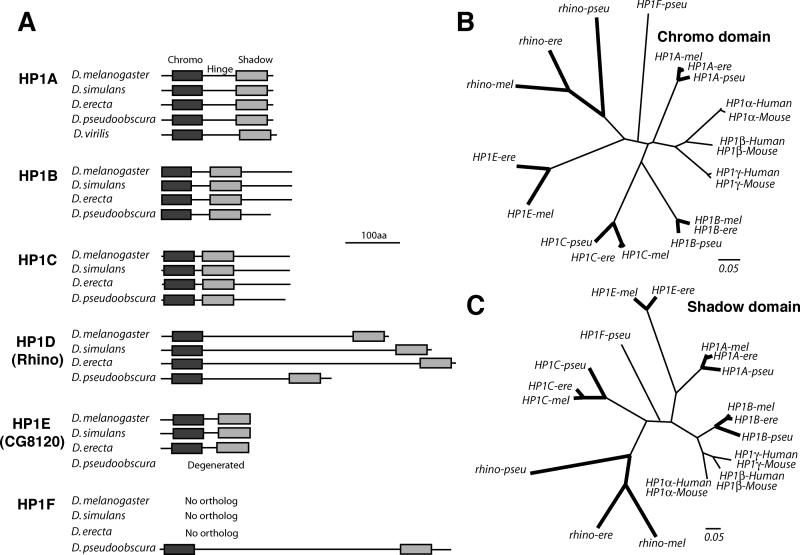
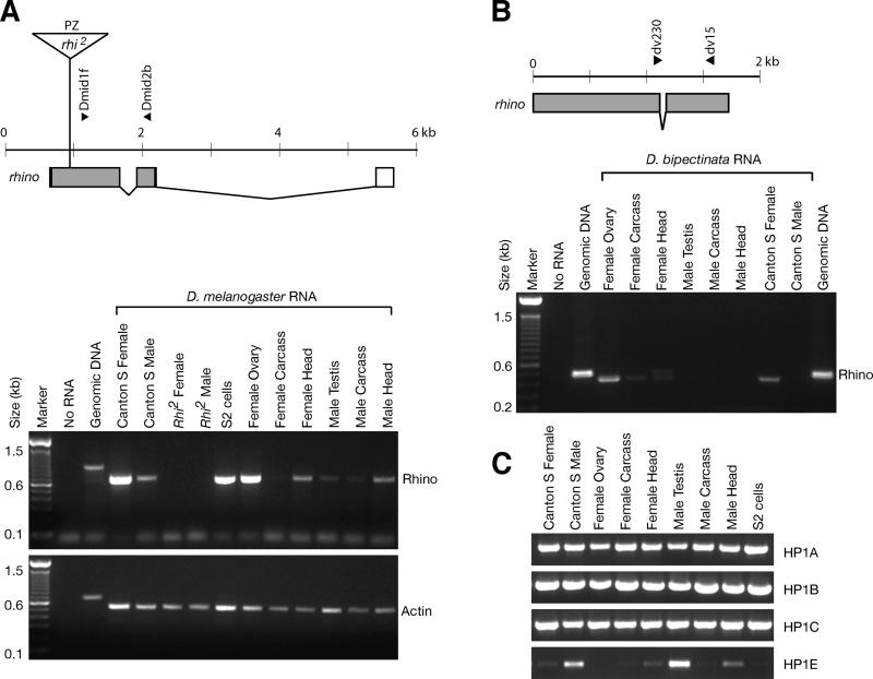
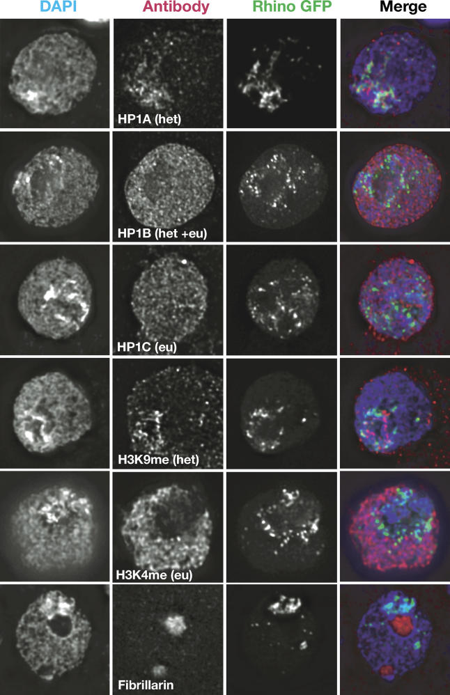
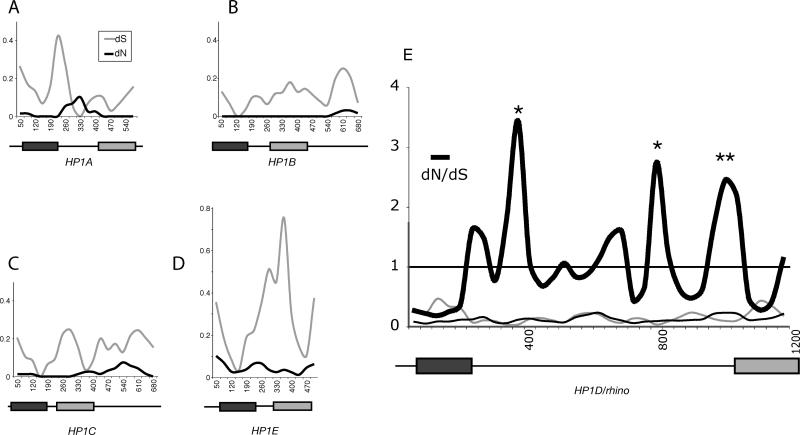
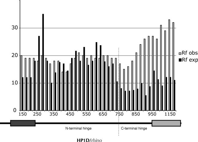
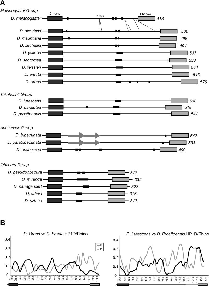
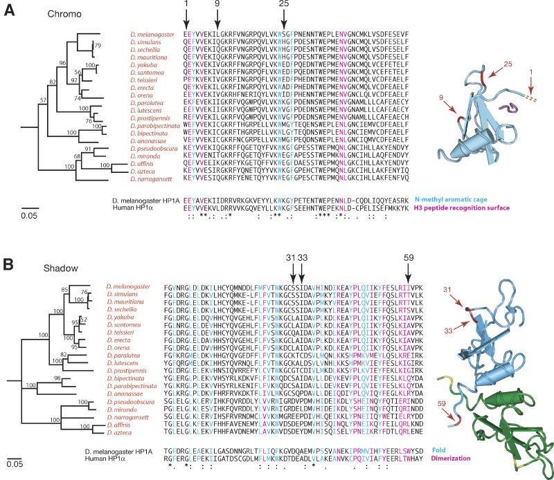
Heterochromatin comprises a significant component of many eukaryotic genomes. In comparison to euchromatin, heterochromatin is gene poor, transposon rich, and late replicating. It serves many important biological roles, from gene silencing to accurate chromosome segregation, yet little is known about the evolutionary constraints that shape heterochromatin. A complementary approach to the traditional one of directly studying heterochromatic DNA sequence is to study the evolution of proteins that bind and define heterochromatin. One of the best markers for heterochromatin is the heterochromatin protein 1 (HP1), which is an essential, nonhistone chromosomal protein. Here we investigate the molecular evolution of five HP1 paralogs present in Drosophila melanogaster. Three of these paralogs have ubiquitous expression patterns in adult Drosophila tissues, whereas HP1D/rhino and HP1E are expressed predominantly in ovaries and testes respectively. The HP1 paralogs also have distinct localization preferences in Drosophila cells. Thus, Rhino localizes to the heterochromatic compartment in Drosophila tissue culture cells, but in a pattern distinct from HP1A and lysine-9 dimethylated H3. Using molecular evolution and population genetic analyses, we find that rhino has been subject to positive selection in all three domains of the protein: the N-terminal chromo domain, the C-terminal chromo-shadow domain, and the hinge region that connects these two modules. Maximum likelihood analysis of rhino sequences from 20 species of Drosophila reveals that a small number of residues of the chromo and shadow domains have been subject to repeated positive selection. The rapid and positive selection of rhino is highly unusual for a gene encoding a chromosomal protein and suggests that rhino is involved in a genetic conflict that affects the germline, belying the notion that heterochromatin is simply a passive recipient of "junk DNA" in eukaryotic genomes.


Conflict of interest statement

Competing interests. The authors have declared that no competing interests exist.

Figures

Figure 1
Figure 1. HP1 Paralogs in Drosophila
(A) Proteins encoded by D. melanogaster HP1s and selected orthologs (obtained by PCR from syntenic locations) are drawn to scale (indicated at bottom) with a dark rectangle resembling the N-terminal chromo domain and a lighter rectangle the C-terminal chromo shadow domain. The HP1E open reading frame is no longer preserved in D. pseudoobscura, and D. melanogaster does not contain HP1F. The hinge regions and N- and C-terminal extensions cannot be aligned between different HP1 types, for example HP1A versus HP1B. HP1D/Rhino contains a very long hinge region that is poorly conserved between species. (B) A neighbor-joining phylogenetic tree based on an alignment of selected HP1 chromo and (C) shadow domains. The monophyletic vertebrate HP1 paralogs are shown for comparison. rhino evolution is clearly distinct from vertebrate or other Drosophila HP1s. HP1 orthologs between D. melanogaster, D. erecta, and D pseudoobscura are shown connected by bold branches (HP1E is not conserved in D. pseudoobscura). The divergence times for D. melanogaster–D. erecta and D. melanogaster–D. pseudoobscura are approximately 9 and 25 million years respectively, whereas those for mouse–human are approximately 80 million years. Clearly, the rhino chromo and shadow domains are far more divergent between these Drosophila species than the chromo domains of HP1A, -B, and -C.
Figure 2
Figure 2. RT-PCR Analyses of the Various HP1 Paralogs
(A) The rhino gene from D. melanogaster is drawn to scale. Exons are boxed (grey fill indicates coding sequence) and lines indicate introns. The position of a P[lacZ, ry+] (PZ) element in the rhi2 mutant is shown (triangle; not to scale). Dmid1f and Dmid2b RT-PCR primers span the first rhino intron. RT-PCR was carried out on roughly equivalent amounts of RNA using a primer set for rhino or actin-42A (primer sequences in Table S2). Control reactions contained no RNA or D. melanogaster genomic DNA. (B) The rhino gene from D. bipectinata is schematized and primers used for RT-PCR indicated. RT-PCR analysis shows that rhino is specifically expressed in ovaries. D. bipectinata separated from D. melanogaster approximately 13 million years ago. (C) RT-PCR reactions carried out for the other HP1 paralogs in D. melanogaster. HP1A, -B, and -C are ubiquitously expressed in adult tissues whereas HP1E expression appears to be predominantly restricted to the male testes.
Figure 3
Figure 3. Rhino-GFP Localizes to Distinct Foci in the Heterochromatic Domain
A C-terminal GFP fusion protein of rhino was transiently expressed in Drosophila tissue culture cells (green in merge). Nuclei were stained with DAPI that stains DNA (blue in merge) and antibodies (red in merge) to HP1A, HP1B, HP1C, H3K9me, H3K4me, or fibrillarin (a nucleolar protein). H3K4me stains euchromatin whereas HP1A, H3K9me, Rhino-GFP, and bright DAPI staining all fall within heterochromatin. Rhino-GFP does not overlap with any of the antibody staining patterns, but appears to localize adjacent to HP1A and H3K9me within the heterochromatic domain.
Figure 4
Figure 4. Comparison of D. melanogaster and D. simulans HP1s
(A–E) Different D. melanogaster and D. simulans HP1 coding DNA sequences were aligned (indels and unalignable sequences were removed) and dN (black line) and dS (grey line) values were calculated using K-estimator [75] with a sliding window of 100 bases and a 35-bp step size. The domain structure of each HP1 is shown schematically and to scale beneath each plot, with the dark rectangle representing the chromo domain and the grey rectangle the chromo shadow domain. For HP1A, dN exceeds dS in the hinge region, but dS is very low in these windows. In contrast, for rhino, dN is higher throughout and exceeds dS in several windows corresponding to the hinge region (dN/dS values are also plotted for rhino). Windows in which statistically significant values for positive selection were obtained (dN/dS > 1, p < 0.02), are indicated by asterisks and map to the hinge region.
Figure 5
Figure 5. Population Genetics of HP1D/rhino between D. melanogaster and D. simulans
Replacement changes that have been fixed between D. melanogaster (17 strains) and D. simulans (11 strains) (Rf obs [observed], open bars) were calculated with a 300-nucleotide sliding window, 25-nucleotide step size. The number of expected replacement changes for each window (Rf exp; solid bars) were calculated from the neutral expectation of the McDonald-Kreitman test (Rf:Sf ≈ Rp:Sp). Rf obs exceeds Rf exp in the C-terminal part of rhino (the C-terminal part of the hinge and the shadow domain as shown beneath), consistent with positive selection (also see Table 1). The chromo and chromo shadow domains are represented by dark and light rectangles, respectively.
Figure 6
Figure 6. Positive Selection of rhino in 25 Million Years of Drosophila Evolution
(A) rhino was PCR amplified and sequenced from the indicated Drosophila species. Predicted protein sequences are drawn to scale with amino acid length shown on the right. The chromo and chromo shadow domains are relatively conserved and are indicated by the large dark and light rectangles, respectively. The hinge regions are rapidly evolving. They differ dramatically in size and sequence and cannot be aligned between different species groups (indicated on the left) and sometimes not even within the same species group, for example the D. bipectinata versus D. ananassae hinge. Within the melanogaster species group, D. melanogaster rhino appears to have undergone large deletions up to 50 codons in the hinge region compared with its closest relative D. simulans (indicated by slanted lines). These deletions are adjacent to the adaptively evolving hinge region identified between D. melanogaster and D. simulans. A 58 amino acid duplication present in the ananassae species group is indicated by grey arrows. Thin black rectangles indicate runs of serine ranging between 70% and 100% serine. (B) dN and dS calculations for rhino from alignments of two pairs of closely related species from the melanogaster and takahashii species groups show multiple windows in which dN exceeds dS, indicative of positive selection.
Figure 7
Figure 7. Positive Selection of the Chromo and Chromo Shadow Domains of rhino
(A) An amino acid alignment of the chromo domain of different Drosophila species is shown with the distantly related HP1A and human HP1α chromo domains for comparison. The neighbor-joining tree based on this alignment (shown on the left) recapitulates known Drosophila phylogeny. Amino acids of the HP1A chromo domain that are involved in binding to H3K9me are color coded: Blue amino acids form an aromatic cage that recognize K9me, and pink amino acids form a complementary surface for recognition of the H3 peptide [45]. The corresponding DNA sequence alignment was used in a PAML analysis. Three codons that have been under repeated and strong positive selection are indicated by arrows. The corresponding positions (red) are indicated on the known structure of the Drosophila HP1A chromo domain (light blue) bound to H3K9me (purple) [45]. (B) Amino acid alignment of representative chromo shadow domains of rhino orthologs from Drosophila. The neighbor-joining tree based on this alignment also recapitulates Drosophila phylogeny. Amino acids of mouse HP1β known to be involved in dimerization are shown in pink and those required for the shadow fold in blue [46]. We use arrows to indicate codons identified as being under positive selection by our PAML analysis. Corresponding positions of the mouse HP1β chromo shadow domain are indicated (red) on one of the shadow domains (light blue) of the dimer [46]. These positions are shown in yellow on the other shadow domain (green).

Similar articles

Cited by

References

    1. Hodgetts R. Eukaryotic gene regulation by targeted chromatin re-modeling at dispersed, middle-repetitive sequence elements. Curr Opin Genet Dev. 2004;14:680–685. - PubMed
    1. Kazazian HH., Jr Mobile elements: Drivers of genome evolution. Science. 2004;303:1626–1632. - PubMed
    1. Deininger PL, Moran JV, Batzer MA, Kazazian HH., Jr Mobile elements and mammalian genome evolution. Curr Opin Genet Dev. 2003;13:651–658. - PubMed
    1. Malik HS, Henikoff S. Conflict begets complexity: The evolution of centromeres. Curr Opin Genet Dev. 2002;12:711–718. - PubMed
    1. Choo KH. Domain organization at the centromere and neocentromere. Dev Cell. 2001;1:165–177. - PubMed