

Synergistic induction of tissue factor by coagulation factor Xa and TNF: evidence for involvement of negative regulatory signaling cascades
- PMID: 16105945
- PMCID: PMC1189324
- DOI: 10.1073/pnas.0504526102
Synergistic induction of tissue factor by coagulation factor Xa and TNF: evidence for involvement of negative regulatory signaling cascades
Abstract
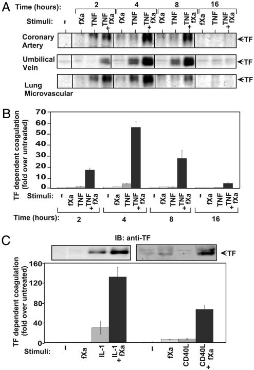
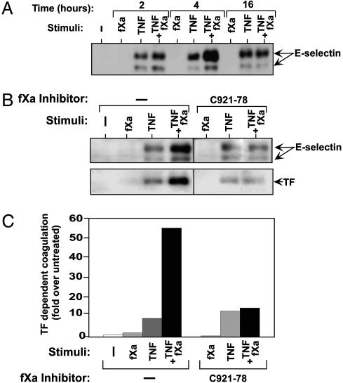
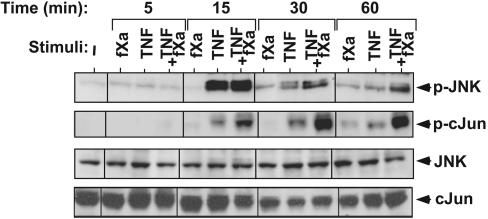
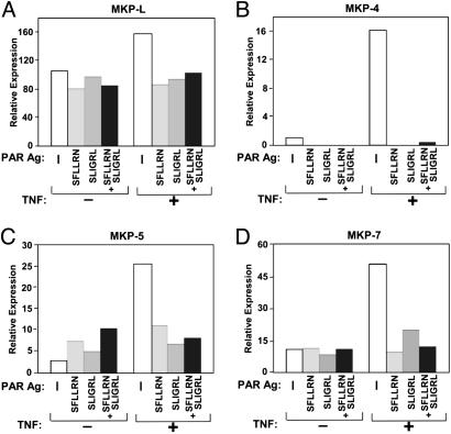
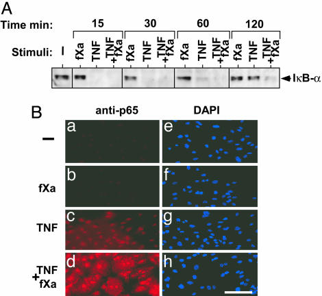
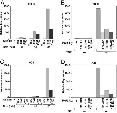
Enzymes of the blood coagulation pathway enhance the inflammatory response leading to endothelial dysfunction, accounting, in part, for the vascular complications occurring in sepsis and cardiovascular disease. The responses of endothelial cell activation include induction of the expression of tissue factor (TF), a membrane glycoprotein that promotes thrombosis, and of E-selectin, a cell adhesion molecule that promotes inflammation. In this report, we demonstrate synergistic interactions between the coagulation factor Xa (fXa) and the proinflammatory cytokines TNF, IL-1beta, and CD40L, leading to enhanced expression of TF and E-selectin in endothelial cells. A detailed analysis of the molecular pathways that could account for this activity of fXa showed that fXa inhibited the cytokine-induced expression of dual specificity phosphatases, MAP kinase phosphatase-L, -4, -5, and -7, blocking a negative regulatory effect on c-Jun N-terminal kinase. The synergistic interaction between fXa and TNF was also involved in the inhibition of A20 and IkappaBalpha expression in the IkappaB kinase-NF-kappaB pathway. The data indicate that inhibition of negative regulatory signaling accounts for the amplification of cytokine-induced endothelial cell activation by fXa.
Figures

Similar articles
-
Adiponectin inhibits tissue factor expression and enhances tissue factor pathway inhibitor expression in human endothelial cells.Thromb Haemost. 2008 Aug;100(2):291-300. Thromb Haemost. 2008. PMID: 18690350
-
ZLJ-6, a novel COX/5-LOX inhibitor, attenuates TNF-α-induced endothelial E-selectin, ICAM-1 and VCAM-1 expression and monocyte-endothelial interactions via a COX/5-LOX-independent mechanism.Vascul Pharmacol. 2011 Nov-Dec;55(5-6):135-42. doi: 10.1016/j.vph.2011.07.003. Epub 2011 Jul 12. Vascul Pharmacol. 2011. PMID: 21777697
-
Effects of antioxidants and NO on TNF-alpha-induced adhesion molecule expression in human pulmonary microvascular endothelial cells.Respir Med. 2005 May;99(5):580-91. doi: 10.1016/j.rmed.2004.10.007. Epub 2004 Nov 18. Respir Med. 2005. PMID: 15823455
-
Blood coagulation factor Xa as an emerging drug target.Expert Opin Ther Targets. 2011 Mar;15(3):341-9. doi: 10.1517/14728222.2011.553608. Epub 2011 Jan 21. Expert Opin Ther Targets. 2011. PMID: 21250873 Review.
-
Non-hemostatic activity of coagulation factor Xa: potential implications for various diseases.Curr Opin Pharmacol. 2001 Apr;1(2):169-75. doi: 10.1016/s1471-4892(01)00033-9. Curr Opin Pharmacol. 2001. PMID: 11714092 Review.
Cited by
-
New prognostic biomarkers of mortality in patients undergoing liver transplantation for hepatocellular carcinoma.World J Gastroenterol. 2018 Oct 7;24(37):4230-4242. doi: 10.3748/wjg.v24.i37.4230. World J Gastroenterol. 2018. PMID: 30310256 Free PMC article. Review.
-
Antiphospholipid Antibodies as Key Players in Systemic Lupus Erythematosus: The Relationship with Cytokines and Immune Dysregulation.Int J Mol Sci. 2024 Oct 20;25(20):11281. doi: 10.3390/ijms252011281. Int J Mol Sci. 2024. PMID: 39457063 Free PMC article. Review.
-
Biomarker Profile of Sepsis-Associated Coagulopathy Using Biochip Assay for Inflammatory Cytokines.Clin Appl Thromb Hemost. 2018 May;24(4):625-632. doi: 10.1177/1076029617709084. Epub 2017 May 17. Clin Appl Thromb Hemost. 2018. PMID: 28514870 Free PMC article.
-
Functional epistatic interaction between rs6046G>A in F7 and rs5355C>T in SELE modifies systolic blood pressure levels.PLoS One. 2012;7(7):e40777. doi: 10.1371/journal.pone.0040777. Epub 2012 Jul 18. PLoS One. 2012. PMID: 22815813 Free PMC article.
-
NDRG1 Signaling Is Essential for Endothelial Inflammation and Vascular Remodeling.Circ Res. 2023 Feb 3;132(3):306-319. doi: 10.1161/CIRCRESAHA.122.321837. Epub 2022 Dec 23. Circ Res. 2023. PMID: 36562299 Free PMC article.
References
Publication types
MeSH terms
Substances
LinkOut - more resources
Full Text Sources
Other Literature Sources
Research Materials
Miscellaneous
