. 2005 Aug 30;102(35):12356-8.
doi: 10.1073/pnas.0503893102. Epub 2005 Aug 16.

Structure of dimeric mitochondrial ATP synthase: novel F0 bridging features and the structural basis of mitochondrial cristae biogenesis

Affiliations

Structure of dimeric mitochondrial ATP synthase: novel F0 bridging features and the structural basis of mitochondrial cristae biogenesis

Fernando Minauro-Sanmiguel et al. Proc Natl Acad Sci U S A. .

Abstract

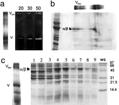
The F1F0-ATP synthase exists as a dimer in mitochondria, where it is essential for the biogenesis of the inner membrane cristae. How two ATP synthase complexes dimerize to promote cristae formation is unknown. Here we resolved the structure of the dimeric F1F0 ATP synthase complex isolated from bovine heart mitochondria by transmission electron microscopy. The structure of the ATP synthase dimer has an overall conic appearance that is consistent with the proposed role of the dimeric enzyme in mitochondrial cristae biogenesis. The ATP synthase dimer interface is formed by contacts on both the F0 and F1 domains. A cross-bridging protein density was resolved which connects the two F0 domains on the intermembrane space side of the membrane. On the matrix side of the complex, the two F1 moieties are connected by a protein bridge, which is attributable to the IF1 inhibitor protein.


Figures

Fig. 1.
Fig. 1.
Gel analysis of dimeric ATP synthase. (a) Digitonin extracts of mitochondrial protein complexes were subjected to BN/PAGE and stained with Coomassie blue (Left) or developed for ATPase activity (Right, containing 20–50 μg of extracted protein). (b) An excised lane from the BN/PAGE was loaded as shown on top of a SDS/polyacrylamide gel together with an enriched F1F0 standard (left lane). Major F1 subunits (α and β) and dimeric F1F0 (Vdim) are indicated. (c) Monomeric (V) and dimeric (Vdim) F1F0 were identified by BN/PAGE in a mitochondrial digitonin extract (Left). The digitonin extract was subsequently loaded on a glycerol density gradient, and fractions obtained after centrifugation were analyzed by SDS/PAGE (Right). Dimeric F1F0 (fraction 7) was resolved from the monomer (fractions 4 and 5). The lane marked WS shows molecular mass standards. For details, see text.
Fig. 2.
Fig. 2.
EM and image analysis of ATP synthase dimer. Dimeric F1F0 enriched as described in the text and in Fig. 1 was used for EM analysis. (a) Area of a typical electron micrograph used for collecting single images. (Scale bar: 50 nm.) The ATP synthase dimers are indicated by boxes. (b) Representative images of the F1F0 dimer. (c) Total average of a data set of 1,130 ATP synthase dimers after the final alignment step. (dh) Averages of the aligned data set after sorting by multivariate statistical analysis. Between 100 and 150 individual images were averaged to produce the images shown. (i) Current working model of the dimeric F1F0 ATP synthase complex. We propose that the association of the two F0 domains involves subunits e and g, which have been shown to be essential for dimer formation. The subunit arrangement is drawn according to cross-linking (16, 23) and EM data (–10). F1 association might result from IF1 bridging in a similar way as seen in the crystal structure of the dimer of soluble F1–ATPase (3). The proposed twofold symmetry axis is indicated by the dotted line. For details, see text.

References

    1. Boyer, P. D. (2000) Biochim. Biophys. Acta 1458, 252–262. - PubMed
    1. Pullman, M. E. & Monroy, G. C. (1963) J. Biol. Chem. 238, 3762–3769. - PubMed
    1. Cabezón, E., Montgomery, M. G., Leslie, A. G. & Walker, J. E. (2003) Nat. Struct. Biol. 10, 744–750. - PubMed
    1. Cabezon, E., Arechaga, I., Jonathan, P., Butler, G. & Walker, J. E. (2000) J. Biol. Chem. 275, 28353–28355. - PubMed
    1. Stock, D., Leslie, A. G. & Walker, J. E. (1999) Science 286, 1687–1688. - PubMed

Publication types

MeSH terms

Substances

LinkOut - more resources