. 2005 Dec 1;106(12):3907-16.
doi: 10.1182/blood-2005-03-1204. Epub 2005 Aug 16.

p130Cas mediates the transforming properties of the anaplastic lymphoma kinase

Affiliations

p130Cas mediates the transforming properties of the anaplastic lymphoma kinase

Chiara Ambrogio et al. Blood. .

Erratum in

  • Blood. 2009 Sep 24;114(13):2851

Abstract

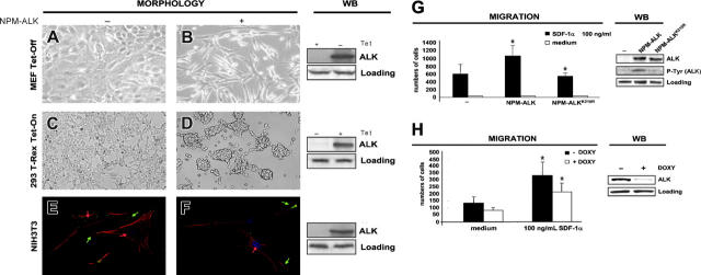
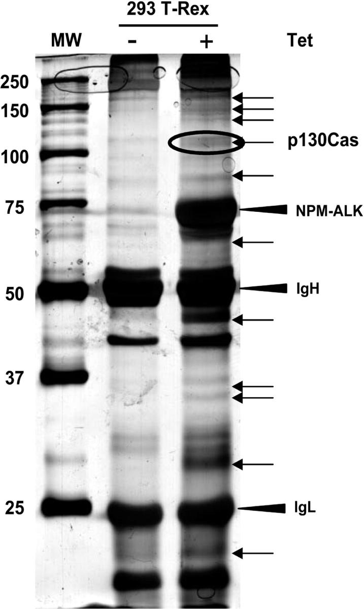
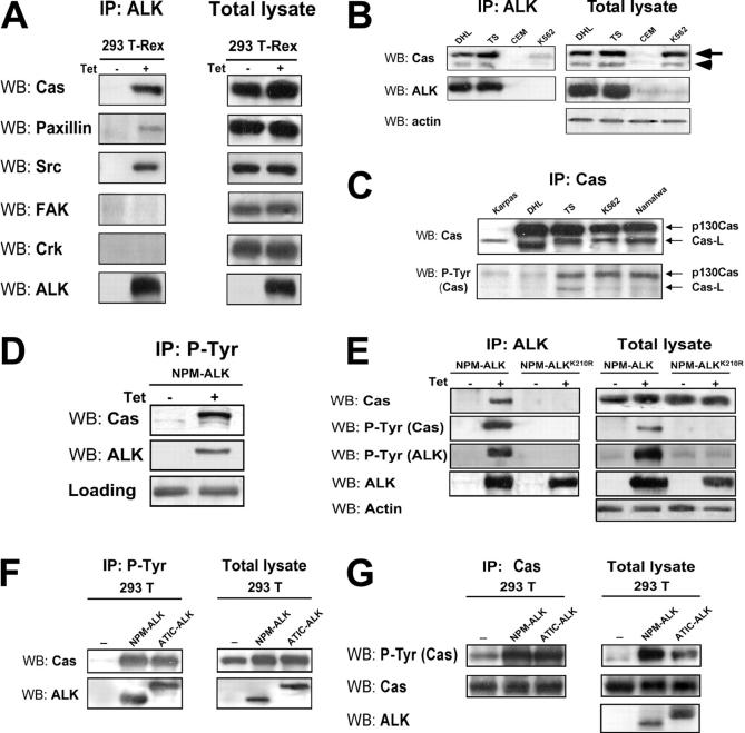
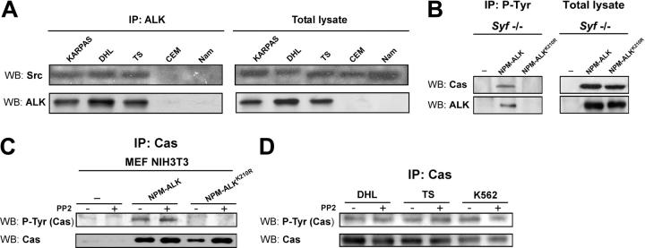
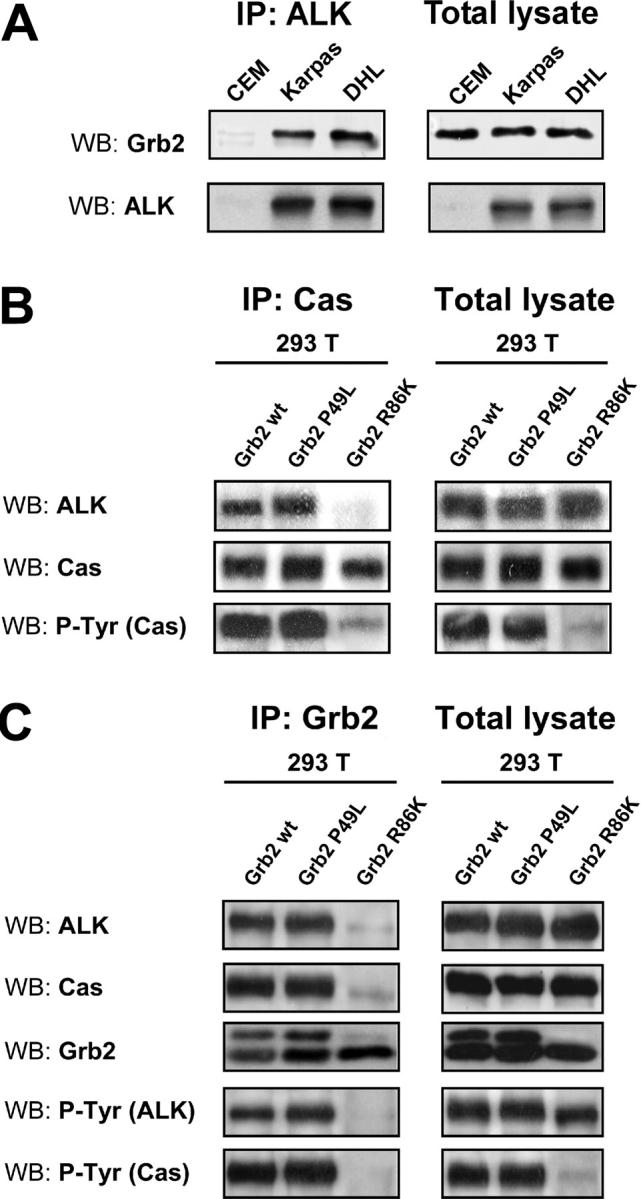
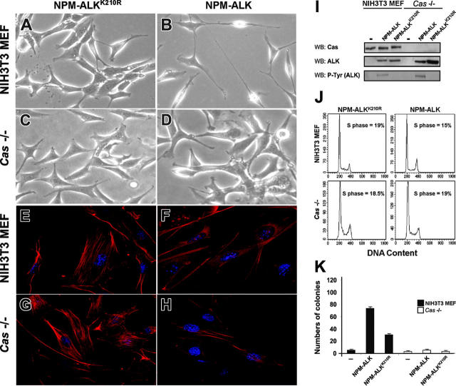
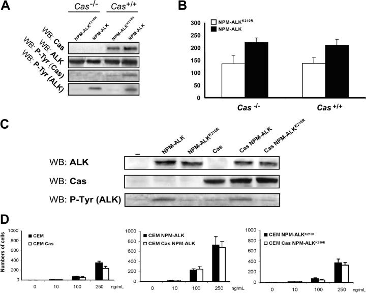
Translocations of the anaplastic lymphoma kinase (ALK) gene have been described in anaplastic large-cell lymphomas (ALCLs) and in stromal tumors. The most frequent translocation, t(2;5), generates the fusion protein nucleophosmin (NPM)-ALK with intrinsic tyrosine kinase activity. Along with transformation, NPM-ALK induces morphologic changes in fibroblasts and lymphoid cells, suggesting a direct role of ALK in cell shaping. In this study, we used a mass-spectrometry-based proteomic approach to search for proteins involved in cytoskeleton remodeling and identified p130Cas (p130 Crk-associated substrate) as a novel interactor of NPM-ALK. In 293 cells and in fibroblasts as well as in human ALK-positive lymphoma cell lines, NPM-ALK was able to bind p130Cas and to induce its phosphorylation. Both of the effects were dependent on ALK kinase activity and on the adaptor protein growth factor receptor-bound protein 2 (Grb2), since no binding or phosphorylation was found with the kinase-dead mutant NPM-ALK(K210R) or in the presence of a Grb2 dominant-negative protein. Phosphorylation of p130Cas by NPM-ALK was partially independent from Src (tyrosine kinase pp60c-src) kinase activity, as it was still detectable in Syf-/- cells. Finally, p130Cas-/- (also known as Bcar1-/-) fibroblasts expressing NPM-ALK showed impaired actin filament depolymerization and were no longer transformed compared with wild-type cells, indicating an essential role of p130Cas activation in ALK-mediated transformation.


Figures

Figure 1.
Figure 1.
Morphologic changes induced by NPM-ALK. (A-B) MEFs Tet-Off show spindle-transformed cell shape when NPM-ALK is expressed. MEFs Tet-Off were stably transfected with the inducible vector NPM-ALK pBIEGFP and forced to express NPM-ALK when grown in medium deprived of tetracycline for 48 hours. (C-D) The 293 T-Rex Tet-On cells undergo changes in adherence when NPM-ALK is induced by adding tetracycline to the culture medium for 24 hours. Contrast-phase images; 40×/0.55 objective lens. (E-F) NIH3T3 fibroblasts were infected with Pallino NPM-ALK retrovirus and then incubated with monoclonal antipaxillin primary antibody followed by FITC-conjugated secondary antibody (green) and with PE-conjugated phalloidin to stain the actin filaments (red). Noninfected NIH3T3 cells (E) show a spread morphology, with clearly detectable actin filaments ending in the focal contacts (red arrows) and with organized paxillin clusters (green arrows), whereas NIH3T3 NPM-ALK cells (F) show only few actin filaments (red arrow) and rare paxillin clusters (green arrows). Images were taken with the Leica confocal microscope using a 63×/1.32 objective lens. Right panels show NPM-ALK expression by WB in the corresponding cells. Samples were blotted with anti-ALK monoclonal antibody. (G) CEM cells were infected with retroviruses expressing NPM-ALK or the kinase-dead mutant NPM-ALKK210R as control and then sorted for GFP expression. The histograms represent the numbers of cells migrated in response to SDF-1α in a transwell assay. The histograms summarize the results of 3 independent experiments using triplicate wells for experimental point. The right panels show NPM-ALK expression and phosphorylation by WB in the corresponding cells. Samples were blotted with anti-ALK and anti-PY20 monoclonal antibodies. (H) NPM-ALK silencing by ALK-shRNA decreases the migration rate of TS cells. TS cells were cotransduced with pLVTH ALK-shRNA and pLV-tTRKRAB vectors to obtain a doxycycline-dependent inducible NPM-ALK silencing. Cells were grown in the presence of doxycycline (1 μg/mL) for 72 hours. The histograms represent the numbers of cells migrated in response to SDF-1α in a transwell assay. The histograms are from 3 independent experiments using triplicate wells for experimental point. The right panels show NPM-ALK expression by WB in the corresponding cells. Samples were blotted with anti-ALK monoclonal antibody. *Statistically significant as analyzed by Student t test.
Figure 2.
Figure 2.
Identification of p130Cas from silver-stained SDS-PAGE. The 293 T-Rex Tet-On cells were transfected with NPM-ALK and cultured for 24 hours in presence or absence of tetracycline. Thirty milligrams of total cell lysate was immunoprecipitated with a mixture of antiphosphotyrosine antibodies (4G10; Upstate Biotechnology; and PY20; Transduction Laboratories) and resolved on 8% SDS-PAGE. The gel was silver stained and the bands present only in the induced sample (+), as indicated by the arrows, were analyzed by mass spectrometry. Molecular marker is shown on the left. The p130Cas was identified from the circled band. See “Materials and methods” for details.
Figure 3.
Figure 3.
NPM-ALK binds and phosphorylates p130Cas. (A) The 293 T-Rex Tet-On cells were cultured with tetracycline for 24 hours; total cell lysate was immunoprecipitated (IP) with anti-ALK monoclonal antibody and blotted with the indicated antibodies by WB. NPM-ALK coprecipitates with p130Cas, paxillin, and Src. (B) NPM-ALK coprecipitates both with p130Cas (arrow) and with Cas-L (arrowhead) in lymphoid cell lines. Total lysates from ALK-positive (DHL, TS) and ALK-negative (CEM, K562) cell lines were immunoprecipitated with anti-ALK monoclonal antibody and blotted with the indicated antibodies. (C) In ALK-positive lymphoid cells, both p130Cas and Cas-L are phosphorylated. Total lysates were immunoprecipitated with anti-p130Cas monoclonal antibody and blotted with the anti-PY20 monoclonal antibody. (D) The adaptor p130Cas is phosphorylated in the presence of NPM-ALK. The 293 T-Rex Tet-On cells expressing NPM-ALK were cultured with tetracycline for 24 hours; cell lysate was immunoprecipitated with anti-PY20 monoclonal antibody and blotted with the indicated antibodies. (E) The binding and the phosphorylation of p130Cas depends on NPM-ALK kinase activity. The 293 T-Rex Tet-On cells expressing the active form of NPM-ALK or the kinase-dead control (NPM-ALKK210R) were cultured with tetracycline for 24 hours; cell lysates were immunoprecipitated with anti-ALK monoclonal antibody and blotted with the indicated antibodies. (F-G) The phosphorylation of p130Cas does not depend on the binding with the NPM portion of NPM-ALK. The 293 T cells were transfected with Pallino p130Cas alone or in combination with Pallino NPM-ALK or Pallino ATIC-ALK. Samples were collected 48 hours after transfection and total lysates were immunoprecipitated and blotted with the indicated antibodies.
Figure 4.
Figure 4.
The phosphorylation of p130Cas by NPM-ALK is independent from Src tyrosine kinase activity. (A) NPM-ALK coprecipitates with Src in lymphoid cells. Total lysates from ALK-positive (Karpas, DHL, TS) and ALK-negative (CEM, Namalwa) cell lines were immunoprecipitated with anti-ALK monoclonal antibody and blotted with anti-Src polyclonal antibody. (B) The NPM-ALK–mediated phosphorylation of p130Cas is Src independent. Syf triple knock-out fibroblasts were cotransfected with Pallino p130Cas and Pallino NPM-ALK or Pallino NPM-ALKK210R as a control. Samples were collected 48 hours after transfection and total lysates were immunoprecipitated with anti-PY20 monoclonal antibody and blotted with the indicated antibodies. (C) Src inhibition does not limit p130Cas phosphorylation. MEF NIH3T3 cells were cotransfected with Pallino p130Cas and Pallino NPM-ALK or Pallino NPM-ALKK210R as a control. Forty-eight hours after transfection, samples were cultivated in the presence of 30 μM PP2 for 1 hour and then collected. The adaptor p130Cas phosphorylation levels were detected by immunoprecipitation with anti-p130Cas monoclonal antibody followed by blotting with anti-PY20 monoclonal antibody. (D) ALK-positive (DHL and TS) and ALK-negative (K562) cells were cultivated in the presence of 30 μM PP2 for 1 hour and then collected. The adaptor p130Cas phosphorylation levels were detected by immunoprecipitation with anti-p130Cas monoclonal antibody followed by blotting with anti-PY20 monoclonal antibody.
Figure 5.
Figure 5.
NPM-ALK binds p130Cas through the SH2 domain of Grb2. (A) NPM-ALK binds Grb2 in lymphoid cell lines. Total lysates were immunoprecipitated with anti-ALK monoclonal antibody and blotted with the indicated antibodies. (B-C) The 293 T cells were transfected with Pallino NPM-ALK, Pallino p130Cas, pRK5 Grb2, and the dominant-negative Grb2 constructs pRK5 P49L (mutated in the SH3 domain) or pRK5 R86K (mutated in the SH2 domain) as indicated. Samples were collected 48 hours after transfection and total lysates were immunoprecipitated with anti-Grb2 polyclonal antibody and blotted with the indicated antibodies. Grb2 R86K was able to disrupt the binding of p130Cas to NPM-ALK (B) as well as p130Cas phosphorylation (C).
Figure 6.
Figure 6.
The adaptor p130Cas is required for NPM-ALK–mediated transformation and actin filaments organization. NIH3T3 MEFs and Cas-/- MEFs were infected with Pallino NPM-ALK or Pallino NPM-ALKK210R retroviruses and sorted to obtain greater than 95% GFP-positive cells. (A-B) The expression of NPM-ALK in NIH3T3 MEFs led to spindle-shape morphology and to the outgrowth of cellular processes. NPM-ALKK210R was used as a control. (C-D) Cas-/- cells infected with Pallino NPM-ALK did not show evident morphologic differences in comparison with the control NPM-ALKK210R. Phase-contrast images acquired with a 40×/0.55 objective lens. (E-H) Cas-/- fibroblasts expressing NPM-ALK retain the organization of the actin cytoskeleton. Compared with NPM-ALK NIH3T3 MEFs that undergo an evident loss of the actin structure (F), NPM-ALK Cas-/- fibroblasts show a phenotype resembling Cas-/- infected with the control NPM-ALKK210R (G-H). Cells were incubated with PE-conjugated phalloidin to stain the actin filaments (red). Images were taken with the Leica confocal microscope equipped with a 63×/1.32 oil immersion objective lens. (I) Protein expression levels were analyzes by WB as indicated. (J) Cell-cycle analysis was performed by DNA content evaluation on cells in logarithmic growth phase. (K) NIH3T3 and Cas-/- MEFs, infected as indicated, were plated in soft agar and cultured for 3 weeks. The histograms represent the average numbers of colonies from the indicated cells and constructs. Data are from 1 of 3 independent experiments, each including triplicates for experimental point.
Figure 7.
Figure 7.
The p130Cas is not required for NPM-ALK–mediated migration. (A) Cas-/- and Cas+/+ rescued fibroblasts were infected with Pallino NPM-ALK retrovirus and then sorted for GFP content. Protein expression levels were verified by WB as indicated. (B) Cells were plated on a Matrigel-coated insert and the number of migrated cells was evaluated after 48 hours. The histograms represent the average number of migrated cells from 3 independent experiments using triplicate wells for experimental point. (C) CEM lymphoblastoid cells were infected with Pallino retroviruses containing NPM-ALK or NPM-ALKK210R together with retrovirus for p130Cas. Protein expression levels were verified by WB as indicated. (D) The histograms represent the average numbers of migrated cells in response to increasing concentrations of SDF-1α as indicated. Data are from 1 of 2 independent experiments. Error bars indicate standard deviation. *Statistically significant analysis as measured by the Student t test.

References

    1. Morris SW, Kirstein MN, Valentine MB, et al. Fusion of a kinase gene, ALK, to a nucleolar protein gene, NPM, in non-Hodgkin's lymphoma. Science. 1994;263: 1281-1284. - PubMed
    1. Borer RA, Lehner CF, Eppenberger HM, Nigg EA. Major nucleolar proteins shuttle between nucleus and cytoplasm. Cell. 1989;56: 379-390. - PubMed
    1. Okuda M. The role of nucleophosmin in centrosome duplication. Oncogene. 2002;21: 6170-6174. - PubMed
    1. Kurki S, Peltonen K, Latonen L, et al. Nucleolar protein NPM interacts with HDM2 and protects tumor suppressor protein p53 from HDM2-mediated degradation. Cancer Cell. 2004;5: 465-475. - PubMed
    1. Chan PK, Chan FY. Nucleophosmin/B23 (NPM) oligomer is a major and stable entity in HeLa cells. Biochim Biophys Acta. 1995;1262: 37-42. - PubMed

Publication types

MeSH terms

Substances