

Not so crystal clear: the structure of the human telomere G-quadruplex in solution differs from that present in a crystal
- PMID: 16106044
- PMCID: PMC1187823
- DOI: 10.1093/nar/gki782
Not so crystal clear: the structure of the human telomere G-quadruplex in solution differs from that present in a crystal
Abstract
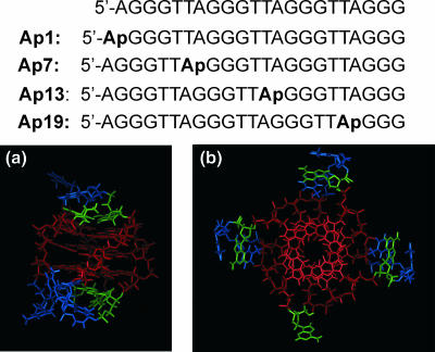
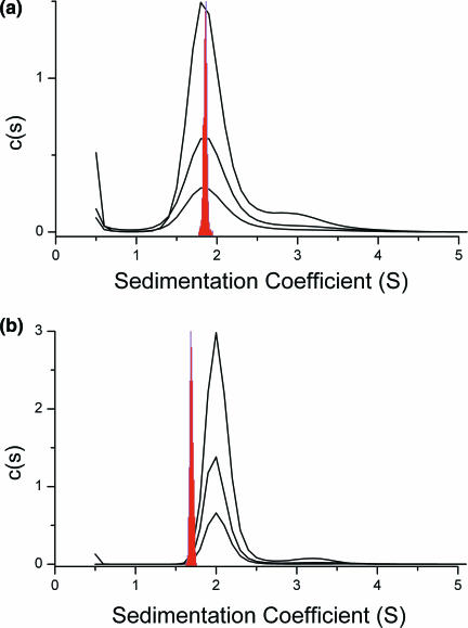
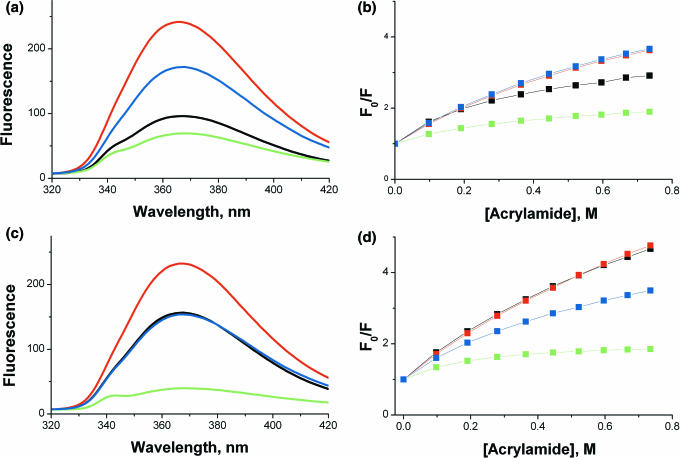
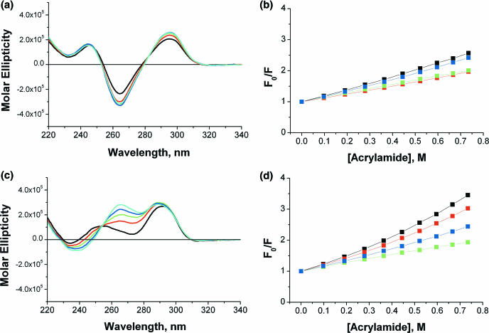
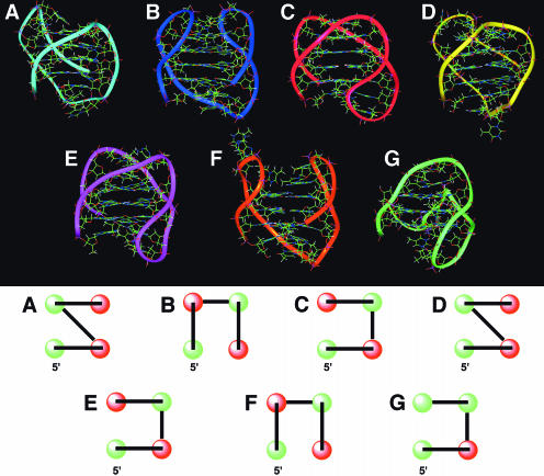
The structure of human telomere DNA is of intense interest because of its role in the biology of both cancer and aging. The sequence [5'-AGGG(TTAGGG)3] has been used as a model for telomere DNA in both NMR and X-ray crystallographic studies, the results of which show dramatically different structures. In Na+ solution, NMR revealed an antiparallel G-quadruplex structure that featured both diagonal and lateral TTA loops. Crystallographic studies in the presence of K+ revealed a flattened, propeller-shaped structure featuring a parallel-stranded G-quadruplex with symmetrical external TTA loops. We report the results of biophysical experiments in solution and computational studies that are inconsistent with the reported crystal structure, indicating that a different structure exists in K+ solutions. Sedimentation coefficients were determined experimentally in both Na+ and K+ solutions and were compared with values calculated using bead models for the reported NMR and crystal structures. Although the solution NMR structure accurately predicted the observed S-value in Na+ solution, the crystal structure predicted an S-value that differed dramatically from that experimentally observed in K+ solution. The environments of loop adenines were probed by quantitative fluorescence studies using strategic and systematic single-substitutions of 2-aminopurine for adenine bases. Both fluorescence intensity and quenching experiments in K+ yielded results at odds with quantitative predictions from the reported crystal structure. Circular dichroism and fluorescence quenching studies in the presence of the crowding agent polyethylene glycol showed dramatic changes in the quadruplex structure in K+ solutions, but not in Na+ solutions, suggesting that the crystal environment may have selected for a particular conformational form. Molecular dynamics simulations were performed to yield model structures for the K+ quadruplex form that are consistent with our biophysical results and with previously reported chemical modification studies. These models suggest that the biologically relevant structure of the human telomere quadruplex in K+ solution is not the one determined in the published crystalline state.
Figures
Similar articles
-
The new models of the human telomere d[AGGG(TTAGGG)3] in K+ solution.Bioorg Med Chem. 2006 Aug 15;14(16):5584-91. doi: 10.1016/j.bmc.2006.04.033. Epub 2006 May 8. Bioorg Med Chem. 2006. PMID: 16682210
-
The new models of the human telomere DNA in K+ solution revealed by NMR analysis assisted by the incorporation of 8-bromoguanines.Nucleic Acids Symp Ser (Oxf). 2006;(50):45-6. doi: 10.1093/nass/nrl023. Nucleic Acids Symp Ser (Oxf). 2006. PMID: 17150809
-
Human telomeric sequence forms a hybrid-type intramolecular G-quadruplex structure with mixed parallel/antiparallel strands in potassium solution.Nucleic Acids Res. 2006 May 19;34(9):2723-35. doi: 10.1093/nar/gkl348. Print 2006. Nucleic Acids Res. 2006. PMID: 16714449 Free PMC article.
-
Diverse effects of naturally occurring base lesions on the structure and stability of the human telomere DNA quadruplex.Biochimie. 2015 Nov;118:15-25. doi: 10.1016/j.biochi.2015.07.013. Epub 2015 Jul 15. Biochimie. 2015. PMID: 26188111 Review.
-
Fluorescence detection of potassium ion using the G-quadruplex structure.Anal Sci. 2011;27(12):1167-72. doi: 10.2116/analsci.27.1167. Anal Sci. 2011. PMID: 22156241 Review.
Cited by
-
Relative stability of different DNA guanine quadruplex stem topologies derived using large-scale quantum-chemical computations.J Am Chem Soc. 2013 Jul 3;135(26):9785-96. doi: 10.1021/ja402525c. Epub 2013 Jun 19. J Am Chem Soc. 2013. PMID: 23742743 Free PMC article.
-
Processing of G4 DNA by Dna2 helicase/nuclease and replication protein A (RPA) provides insights into the mechanism of Dna2/RPA substrate recognition.J Biol Chem. 2008 Sep 5;283(36):24359-73. doi: 10.1074/jbc.M802244200. Epub 2008 Jun 30. J Biol Chem. 2008. PMID: 18593712 Free PMC article.
-
Structure-based drug design: from nucleic acid to membrane protein targets.Exp Mol Pathol. 2009 Jun;86(3):141-50. doi: 10.1016/j.yexmp.2009.01.011. Epub 2009 Jan 31. Exp Mol Pathol. 2009. PMID: 19454265 Free PMC article. Review.
-
Mechanical unfolding of human telomere G-quadruplex DNA probed by integrated fluorescence and magnetic tweezers spectroscopy.Nucleic Acids Res. 2013 Feb 1;41(4):2746-55. doi: 10.1093/nar/gks1341. Epub 2013 Jan 8. Nucleic Acids Res. 2013. PMID: 23303789 Free PMC article.
-
Thermodynamic characterization of human telomere quadruplex unfolding.Biopolymers. 2013 Dec;99(12):1006-18. doi: 10.1002/bip.22247. Biopolymers. 2013. PMID: 23536479 Free PMC article.
References
-
- Hurley L.H. DNA and its associated processes as targets for cancer therapy. Nature Rev. Cancer. 2002;2:188–200. - PubMed
-
- Mergny J.L., Helene C. G-quadraplex DNA: A target for drug design. Nature Medicine. 1998;4:1366–1367. - PubMed
-
- Neidle S., Read M.A. G-quadruplexes as therapeutic targets. Biopolymers. 2000;56:195–208. - PubMed
