Comparative Study
. 2005 Aug 17;25(33):7481-8.
doi: 10.1523/JNEUROSCI.1801-05.2005.

Strong effects of subphysiological temperature on the function and plasticity of mammalian presynaptic terminals

Affiliations
Comparative Study

Strong effects of subphysiological temperature on the function and plasticity of mammalian presynaptic terminals

Kristina D Micheva et al. J Neurosci. .

Abstract

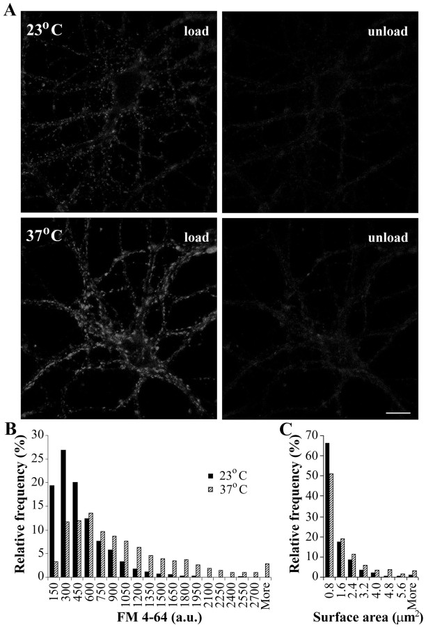
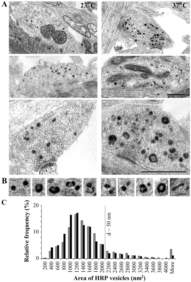
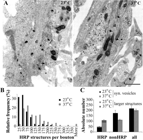
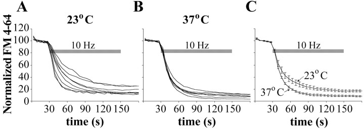
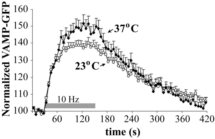
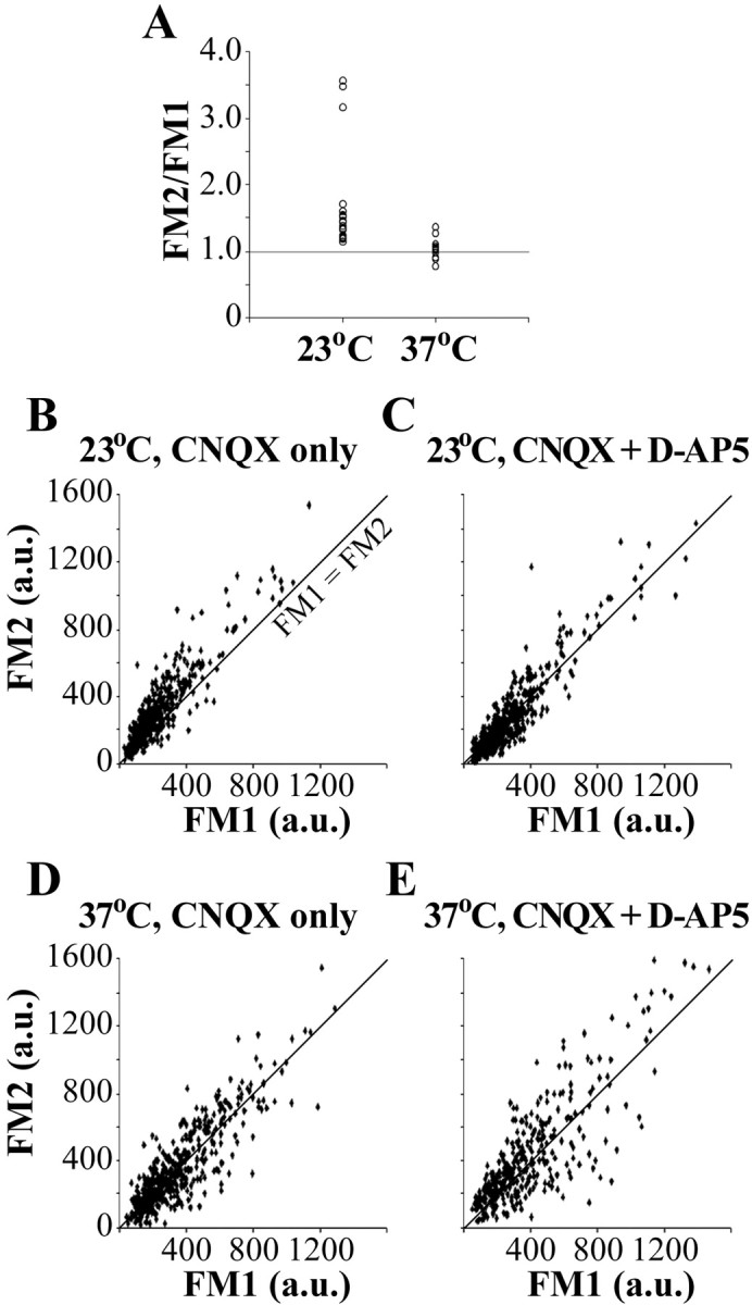
Most cellular processes are known to be strongly temperature dependent. Nevertheless, a large fraction of studies of mammalian synaptic function have been and are performed near room temperature (i.e., at least 10 degrees C below physiological temperature). Here, we examined the effects of temperature on presynaptic function in primary cultures of rat hippocampal neurons. FM dyes, VAMP (vesicle-associated membrane protein)-GFP (green fluorescent protein) transfection, and HRP uptake were used to quantify various aspects of synaptic vesicle recycling. Our results show that there are very substantial differences in synaptic vesicle recycling at physiological temperature as opposed to the common, lower experimental temperatures. At 37 degrees C, compared with 23 degrees C, the speed of both exocytosis and endocytosis was higher. The size of the recycling vesicle pool (in both number of vesicles and spatial extent) was twofold larger at 37 degrees C. In addition, although repeated 10 Hz electrical stimulation caused an NMDA receptor-dependent enlargement (averaging 170%) of the measurable recycling vesicle pool at 23 degrees C, the same stimulus repetition had no effect at 37 degrees C. These results show that it is potentially misleading to extend conclusions drawn about vesicle function or presynaptic plasticity at lowered experimental temperature to physiological conditions and that much new experimental work at the higher physiological temperature range will be needed to understand the true parameters of presynaptic functions.


Figures

Figure 1.
Figure 1.
FM 4-64 loading of hippocampal neurons in culture. A, Confocal images of hippocampal neurons in primary culture (12 d in vitro) loaded with FM 4-64 (left) and subsequently unloaded (right). The neurons were kept at either room temperature (top) or physiological temperature (bottom). Loading was achieved using electrical stimulation at 10 Hz for 1 min and unloading at 10 Hz for 2 min. Scale bar, 10 μm. B, Histogram presenting the distribution of the fluorescence intensity of the FM 4-64 labeling at individual synapses at both temperatures; fluorescence values are corrected for temperature quenching (n = 1281 boutons from 10 coverslips at 23°C, and n = 941 boutons from 9 coverslips at 37°C). C, Distribution of the surface area of individual FM4-64 puncta at both temperatures (n = 673 boutons from 2 coverslips at 23°C, and n = 634 boutons from 2 coverslips at 37°C).
Figure 2.
Figure 2.
Morphology of HRP-labeled structures after electrical stimulation. A, Electron micrographs of presynaptic boutons formed by hippocampal neuronal cultures (15 d in vitro) electrically stimulated in the presence of HRP at 23°C and 37°C. HRP uptake is identified by the dark DAB reaction product. Scale bar, 0.5 μm. B, Examples of HRP-labeled structures at the two temperatures. Scale bar, 100 nm. C, Distribution of the area of HRP vesicles (in square nanometers) as measured on single sections (n = 603 vesicles at 23°C, and n = 610 vesicles at 37°C). d, Diameter.
Figure 3.
Figure 3.
Recycling vesicle pool in hippocampal neurons identified by HRP uptake. A, At 23°C, compared with 37°C, a smaller number of HRP vesicles is observed, and some presynaptic profiles are even entirely devoid of HRP vesicles (asterisk). The arrows indicate presynaptic boutons. d, Dendrite. Scale bar, 0.5 μm. B, Distribution of the number of HRP structures per presynaptic bouton, as determined from serial sections. C, Absolute numbers of HRP, non-HRP, and all vesicles per presynaptic bouton (mean ± SE; n = 82 boutons from 2 coverslips at 23°C, and n = 89 boutons from 2 coverslips at 37°C). syn., Synaptic. Error bars represent SEM.
Figure 4.
Figure 4.
Speed of exocytosis as measured with FM 4-64. A, B, Unloading curves from experiments performed at 23°C (A; n = 8 coverslips with at least 100 boutons each) and 37°C (B; n = 7 coverslips with at least 100 boutons each). FM 4-64 fluorescence intensities are normalized to the initial resting state of each presynaptic bouton and averaged for each experiment. SEs are not presented in A and B, because they were on the order of 1%. C, Comparison of the mean unloading curves from the experiments in A and B with SEs.
Figure 5.
Figure 5.
Speed of endocytosis determined with VAMP-GFP. VAMP-GFP fluorescence changes in presynaptic boutons during electrical stimulation at 23 and 37°C are shown. Mean values of the normalized VAMP-GFP fluorescence intensities and SEs are shown (for 23°C n = 222 boutons from 7 coverslips and 37°C, n = 157 boutons from 7 coverslips).
Figure 6.
Figure 6.
Presynaptic potentiation of hippocampal neurons. A, Synaptic boutons were loaded twice serially with the same stimulation protocol. Mean values from the second FM load normalized to the first load are presented (n = 16 coverslips at 23°C and n = 13 coverslips at 37°C). B-E, Temperature and NMDA receptor dependence of the presynaptic potentiation. Second FM load versus first FM load for individual presynaptic boutons stimulated at 23 or 37°C, in the presence or absence of an NMDA blocker (d-AP-5; 50 μm), is shown. For each plot, n ≥ 150 boutons from two coverslips. The line represents FM1 = FM2. Potentiation is evident only at 23°C in the absence of d-AP-5, where most of the data points are above the FM1 = FM2 line.

References

    1. Aravanis AM, Pyle JL, Tsien RW (2003) Single synaptic vesicles fusing transiently and successively without loss of identity. Nature 423: 643-647. - PubMed
    1. Betz WJ, Mao F, Bewick GS (1992) Activity-dependent fluorescent staining and destaining of living motor nerve terminals. J Neurosci 12: 363-375. - PMC - PubMed
    1. Borst JG, Sakmann B (1998) Calcium current during a single action potential in a large presynaptic terminal of the rat brainstem. J Physiol (Lond) 506: 143-157. - PMC - PubMed
    1. Cochilla AJ, Angleson JK, Betz WJ (1999) Monitoring secretory membrane with FM1-43 fluorescence. Annu Rev Neurosci 22: 1-10. - PubMed
    1. David G, Barrett EF (2000) Stimulation-evoked increases in cytosolic [Ca2+] in mouse motor nerve terminals are limited by mitochondrial uptake and are temperature dependent. J Neurosci 20: 7290-7296. - PMC - PubMed

Publication types

LinkOut - more resources