Comparative Study
. 2005 Aug 17;25(33):7682-6.
doi: 10.1523/JNEUROSCI.2211-05.2005.

A calcium flux is required for circadian rhythm generation in mammalian pacemaker neurons

Affiliations
Comparative Study

A calcium flux is required for circadian rhythm generation in mammalian pacemaker neurons

Gabriella B Lundkvist et al. J Neurosci. .

Abstract

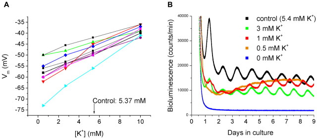
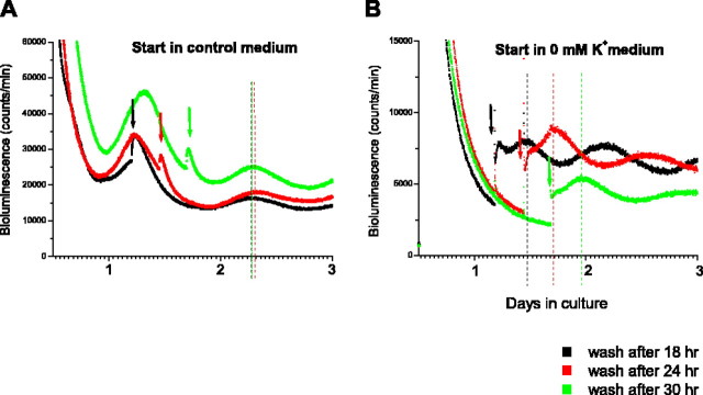
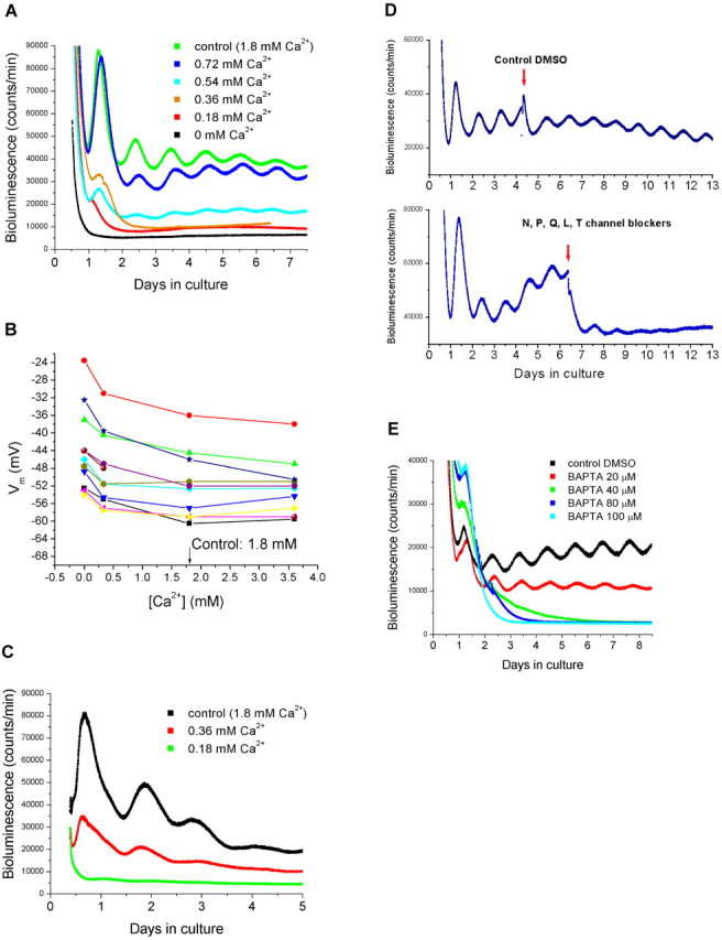
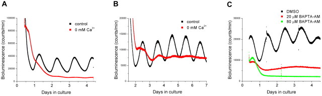
Generation of mammalian circadian rhythms involves molecular transcriptional and translational feedback loops. It is not clear how membrane events interact with the intracellular molecular clock or whether membrane activities are involved in the actual generation of the circadian rhythm. We examined the role of membrane potential and calcium (Ca2+) influx in the expression of the circadian rhythm of the clock gene Period 1 (Per1) within the rat suprachiasmatic nucleus (SCN), the master pacemaker controlling circadian rhythmicity. Membrane hyperpolarization, caused by lowering the extracellular concentration of potassium or blocking Ca2+ influx in SCN cultures by lowering [Ca2+], reversibly abolished the rhythmic expression of Per1. In addition, the amplitude of Per1 expression was markedly decreased by voltage-gated Ca2+ channel antagonists. A similar result was observed for mouse Per1 and PER2. Together, these results strongly suggest that a transmembrane Ca2+ flux is necessary for sustained molecular rhythmicity in the SCN. We propose that periodic Ca2+ influx, resulting from circadian variations in membrane potential, is a critical process for circadian pacemaker function.


Figures

Figure 1.
Figure 1.
SCN recordings of membrane potential and bioluminescence of Per1-luc expression in various levels of [K+]. A, Current-clamp recordings in culture medium containing 0.5, 2.7, 5.4 (control), or 10 mm K+. Each color represents membrane potential in a single neuron in various levels of [K+]. B, Bioluminescence recordings from explants obtained from transgenic Per1-luc rats. The explants were cultured in 0, 0.5, 1, 3, or 5.4 mm K+.
Figure 2.
Figure 2.
Example of SCN phase delays in hyperpolarizing medium. A, Bioluminescence recordings from three explants in control medium represented by green, red, and black traces. The control medium was replaced with fresh control medium at 6 h intervals the second day in culture (indicated by arrows), and the phases were compared using the first peaks after medium replacement as reference points (indicated by dashed lines). B, Bioluminescence recordings from three bilateral explants in medium containing 0 mm K+, represented by green, red, and black traces. The 0 [K+] medium was replaced with control medium at 6 h intervals the second day in culture (indicated by arrows). Dotted lines indicate the peaks of the Per1-luc signals after medium replacement.
Figure 3.
Figure 3.
Recordings of membrane potential and bioluminescence in various concentrations of Ca2+. A, Bioluminescence recordings from SCN explants obtained from transgenic Per1-luc rats. The explants were cultured in medium containing 0, 0.18, 0.36, 0.54, 0.72, or 1.8 mm (control) Ca2+. B, Current-clamp recordings in recording solutions containing 0, 0.33, 1.8, and 3.36 mm Ca2+. Each color represents recorded membrane potentials in a single SCN neuron in various concentrations of Ca2+. C, Bioluminescence recordings from liver explants obtained from Per1-luc rats. Liver tissues were cultured in medium containing 0.18, 0.36, or 1.8 mm Ca2+. D, Effect of Ca2+ channel antagonists on the SCN Per1-luc rhythm. SCN explants were cultured in control medium. After 3-5 d in culture, a DMSO control (top trace) or a mixture of Ca2+ channel blockers specific to N-, P-, Q-, L-, and T-types (bottom trace) was added (indicated by arrows) to the explants. E, The intracellular Ca2+ chelator BAPTA-AM (20, 40, 80, and 100 μm) was added to SCN explants, and bioluminescence was recorded. DMSO was added as a control.
Figure 4.
Figure 4.
Bioluminescence recordings in mouse SCN explants. A, Recordings of Per1-luc signals. The explants were cultured in 0 or 1.8 mm (control) Ca2+. B, Recordings of PER2:LUC expression. Explants were cultured in 0 or 1.8 mm Ca2+. C, Recordings of PER2:LUC expression. Explants were cultured in DMSO control (0.3%) and BAPTA-AM (20 or 80 μm).

References

    1. Abe M, Herzog ED, Yamazaki S, Straume M, Tei H, Sakaki Y, Menaker M, Block GD (2002) Circadian rhythms in isolated brain regions. J Neurosci 22: 350-356. - PMC - PubMed
    1. Aton SJ, Colwell CS, Harmar AJ, Waschek J, Herzog ED (2005) Vasoactive intestinal polypeptide mediates circadian rhythmicity and synchrony in mammalian clock neurons. Nat Neurosci 8: 476-483. - PMC - PubMed
    1. Block GD, Khalsa SB, McMahon DG, Michel S, Geusz M (1993) Biological clocks in the retina: cellular mechanism of biological timekeeping. Int Rev Cytol 146: 83-144. - PubMed
    1. Brereton HM, Harland ML, Froscio M, Petronijevic T, Barritt GJ (1997) Novel variants of voltage-operated calcium channel alpha 1-subunit transcripts in a rat liver-derived cell line: deletion in the IVS4 voltage sensing region. Cell Calcium 22: 39-52. - PubMed
    1. Colwell CS (2001) NMDA-evoked calcium transients and currents in the suprachiasmatic nucleus: gating by the circadian system. Eur J Neurosci 13: 1420-1428. - PMC - PubMed

Publication types

LinkOut - more resources