. 2005 Sep;25(17):7423-31.
doi: 10.1128/MCB.25.17.7423-7431.2005.

p53-Dependent transcriptional repression of c-myc is required for G1 cell cycle arrest

Affiliations

p53-Dependent transcriptional repression of c-myc is required for G1 cell cycle arrest

Jenny S L Ho et al. Mol Cell Biol. 2005 Sep.

Abstract

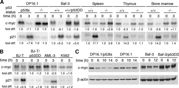
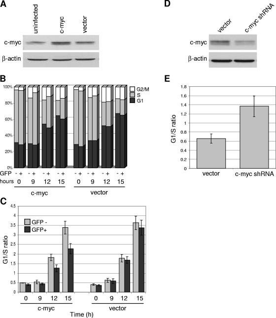
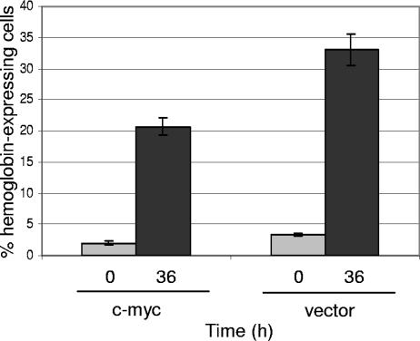
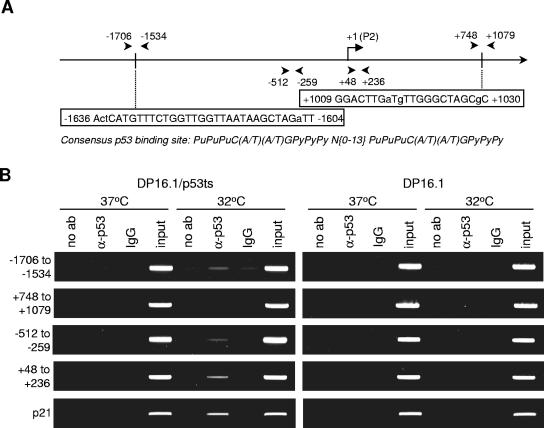
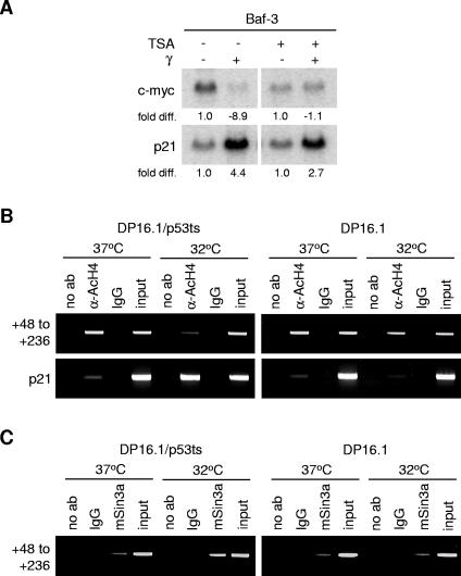
The ability of p53 to promote apoptosis and cell cycle arrest is believed to be important for its tumor suppression function. Besides activating the expression of cell cycle arrest and proapoptotic genes, p53 also represses a number of genes. Previous studies have shown an association between p53 activation and down-regulation of c-myc expression. However, the mechanism and physiological significance of p53-mediated c-myc repression remain unclear. Here, we show that c-myc is repressed in a p53-dependent manner in various mouse and human cell lines and mouse tissues. Furthermore, c-myc repression is not dependent on the expression of p21(WAF1). Abrogating the repression of c-myc by ectopic c-myc expression interferes with the ability of p53 to induce G(1) cell cycle arrest and differentiation but enhances the ability of p53 to promote apoptosis. We propose that p53-dependent cell cycle arrest is dependent not only on the transactivation of cell cycle arrest genes but also on the transrepression of c-myc. Chromatin immunoprecipitation assays indicate that p53 is bound to the c-myc promoter in vivo. We report that trichostatin A, an inhibitor of histone deacetylases, abrogates the ability of p53 to repress c-myc transcription. We also show that p53-mediated transcriptional repression of c-myc is accompanied by a decrease in the level of acetylated histone H4 at the c-myc promoter and by recruitment of the corepressor mSin3a. These data suggest that p53 represses c-myc transcription through a mechanism that involves histone deacetylation.


Figures

FIG. 1.
FIG. 1.
c-myc is repressed in a p53-dependent manner. (A) c-myc transcript is repressed in mouse cells and tissues in a p53-dependent manner. DP16.1/p53ts and DP16.1 cells were maintained at 37°C or shifted to 32°C. Baf-3 and Baf-3/p53DD cells, as well as p53+/+ and p53−/− mice, were left untreated or γ-irradiated (6 Gy). At 3 h after treatment, total RNA was harvested from the cells or mouse tissues (spleen, thymus, and bone marrow). Northern blot analyses were performed, and the blots were hybridized with 32P-labeled cDNA probes for c-myc, p21, and GAPDH (loading control). Signal intensities were quantified by phosphorimaging analyses. Each signal (c-myc or p21) was first normalized to the GAPDH signal within the same sample. In each cell line or tissue, the untreated sample (37°C or unirradiated) signal was assigned a value of 1.0 and the treated sample (32°C or γ-irradiated) was expressed as the fold difference relative to it. (B) p53-dependent repression of c-myc transcript in human cells. BJ-T, BJ-T/p53DD, AML-3, and K562 cells were left untreated or γ-irradiated (6 Gy). Total RNA was harvested from the cells 3 h after treatment. Northern blot analyses and signal quantifications were performed as described for panel A. (C) Western blot analyses using antibodies against c-myc (N-262; Santa Cruz) and β-actin (loading control). DP16.1/p53ts and DP16.1 cells were shifted to 32°C, and Baf-3 and Baf-3/p53DD cells were γ-irradiated (6 Gy). Proteins extracts were made at the times indicated.
FIG. 2.
FIG. 2.
c-myc repression is not mediated by p21. Total RNA was harvested from the thymus, spleen, and bone marrow of p21−/− mice after exposure to γ-irradiation (6 Gy) and subjected to Northern blot analysis as described in the legend for Fig. 1.
FIG. 3.
FIG. 3.
c-myc repression is required for the efficient induction of G1 cell cycle arrest by p53. (A) DP16.1/p53ts cells were retrovirally infected with the pBabe-c-myc-IRES-GFP (c-myc) or pBabe-IRES-GFP (vector) construct. At 48 h after infection, cells were harvested, and protein extracts were prepared and analyzed by Western blotting using an anti-c-myc (N-262; Santa Cruz) or anti-β-actin antibody. Infection efficiencies ranged from 40% to 60%. (B) Cell cycle analyses of cells expressing ectopic c-myc or vector only. DP16.1/p53ts cells infected with c-myc or empty vector (as described for panel A) were shifted to 32°C for the indicated amounts of time. To measure DNA content, cells were stained with Hoechst 33342 and analyzed by flow cytometry to determine the proportion of GFP (uninfected) and GFP+ (infected) cells that were in the G1, S, and G2/M phases of the cell cycle. (C) G1/S ratios were calculated from the data in panel B to portray the extent of G1 cell cycle arrest. Each bar represents the average of three independent experiments ± the standard error. (D) DP16.1/p53ts cells were retrovirally infected with a c-myc shRNA construct or empty vector. At 24 h after infection, cells were harvested, and protein extracts were analyzed by Western blotting. Infection efficiencies ranged from 50% to 70%. (E) Cell cycle analyses of cells expressing c-myc shRNA or vector only. At 24 h after infection, DNA content was determined as described for panel B, and G1/S ratios were determined. Cells were maintained at 37°C throughout. Each bar represents the average of three independent experiments ± the standard error.
FIG. 4.
FIG. 4.
Ectopic c-myc expression interferes with p53-induced cellular differentiation. DP16.1/p53ts cells infected with c-myc or empty vector were incubated at 32°C for 0 or 36 h. Cells were stained for hemoglobin expression using DAF as described elsewhere (16). Stained cells were counted with a hemocytometer, and the proportion of hemoglobin-expressing cells was determined (number of blue cells/total number of cells). Results represent the means of three independent experiments ± the standard errors. Unsorted whole cell populations were used; infection efficiencies ranged from 40 to 60%.
FIG. 5.
FIG. 5.
Ectopic c-myc expression enhances apoptosis induction by p53. DP16.1/p53ts cells infected with c-myc or empty vector were incubated at 32°C for the indicated amounts of time. Cells for the zero-hour time point were maintained at 37°C for the duration of the experiment. Cells were stained with Annexin V-phycoerythrin and 7-AAD and analyzed by flow cytometry to determine the proportion of GFP and GFP+ cells that were apoptotic (Annexin V positive, 7-AAD negative). Each bar represents the mean of three independent experiments ± the standard error.
FIG. 6.
FIG. 6.
p53 binds to the c-myc gene in vivo. (A) Schematic representation of the murine c-myc promoter and 5′ region. The reference point +1 is placed at the transcription start site of the P2 promoter. Arrows indicate the positions of PCR primers used in panel B. Two consensus p53 binding sites and their positions on the gene are also shown; capital letters indicate the residues that satisfy the consensus p53 binding sequence, while lowercase letters indicate mismatches. (B) DP16.1/p53ts and DP16.1 cells were maintained at 37°C or 32°C for 3 h. Cells were harvested for chromatin immunoprecipitation assays as described in Materials and Methods. Samples were immunoprecipitated with no antibody (no ab), a p53 antibody, or rabbit IgG; input controls (1/100 dilution) were also included. PCR analyses on the immunoprecipitated chromatin were carried out using primers that flank the regions as shown in panel A. Numbers on the left of the figure indicate the regions on the c-myc gene that are flanked and amplified by the primers. PCR amplification using primers against a p53 binding site at the p21 promoter was also included as a control.
FIG. 7.
FIG. 7.
p53-mediated transcriptional repression of c-myc involves histone deacetylation at the c-myc promoter. (A) Baf-3 cells were treated with 12.5 nM TSA for 9 h. At 6 h after drug treatment, cells were γ-irradiated where indicated. RNA was harvested 3 h later (9 h total TSA treatment time) and subjected to Northern blot analysis as described in the legend for Fig. 1. (B) Chromatin immunoprecipitations were performed as described in the legend for Fig. 6. Protein-DNA complexes were immunoprecipitated with no antibody (no ab), an antibody against acetylated histone H4 proteins (α-AcH4), or rabbit IgG. (C) Chromatin immunoprecipitations with no antibody (no ab), anti-mSin3a, or rabbit IgG in DP16.1/p53ts and DP16.1 cells.

References

    1. Chen, C. R., Y. Kang, P. M. Siegel, and J. Massague. 2002. E2F4/5 and p107 as Smad cofactors linking the TGFβ receptor to c-myc repression. Cell 110:19-32. - PubMed
    1. Chiou, S. K., L. Rao, and E. White. 1994. Bcl-2 blocks p53-dependent apoptosis. Mol. Cell. Biol. 14:2556-2563. - PMC - PubMed
    1. Coppola, J. A., and M. D. Cole. 1986. Constitutive c-myc oncogene expression blocks mouse erythroleukaemia cell differentiation but not commitment. Nature 320:760-763. - PubMed
    1. D'Souza, S., H. Xin, S. Walter, and D. Choubey. 2001. The gene encoding p202, an interferon-inducible negative regulator of the p53 tumor suppressor, is a target of p53-mediated transcriptional repression. J. Biol. Chem. 276:298-305. - PubMed
    1. Eberhardy, S. R., C. A. D'Cunha, and P. J. Farnham. 2000. Direct examination of histone acetylation on Myc target genes using chromatin immunoprecipitation. J. Biol. Chem. 275:33798-33805. - PubMed

Publication types

MeSH terms