. 2005 Sep;25(17):7616-24.
doi: 10.1128/MCB.25.17.7616-7624.2005.

Structural characterization of the histone variant macroH2A

Affiliations

Structural characterization of the histone variant macroH2A

Srinivas Chakravarthy et al. Mol Cell Biol. 2005 Sep.

Abstract

macroH2A is an H2A variant with a highly unusual structural organization. It has a C-terminal domain connected to the N-terminal histone domain by a linker. Crystallographic and biochemical studies show that changes in the L1 loop in the histone fold region of macroH2A impact the structure and potentially the function of nucleosomes. The 1.6-A X-ray structure of the nonhistone region reveals an alpha/beta fold which has previously been found in a functionally diverse group of proteins. This region associates with histone deacetylases and affects the acetylation status of nucleosomes containing macroH2A. Thus, the unusual domain structure of macroH2A integrates independent functions that are instrumental in establishing a structurally and functionally unique chromatin domain.


Figures
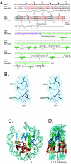
FIG. 1.
FIG. 1.
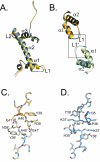
The overall structure of macro-NCP is similar to that of major NCP. (A) Sequence alignment of Xenopus laevis H2A, mouse H2A, and full-length human macroH2A. Filled circles indicate intervals of 10 amino acids in major H2A. Open circles indicate intervals of 10 amino acids in macroH2A. Differences between major H2A (mouse and X. laevis or macroH2A and Xla-H2A, respectively) and macroH2A are shown in red. Differences between mouse and Xla-H2A are shown in blue. The linker region and the nonhistone region of macroH2A are shown in violet and green, respectively. The secondary structure elements of the histone fold (α1, α2, and α3) and extensions (αN and αC) are indicated, as are secondary structure elements for the nonhistoneregion. The fine broken line delineates the docking domain. (B) Stereo view of a section of the  2Fo-Fc  electron density map, calculated at 3Å and contoured at 1σ, showing sequence differences between macroH2A and Xla-H2A L83 to I80, Q84 to L81, and R88 to A85. (C) Superposition of major NCP and macro-NCP (only histone octamers are superimposed) viewed down the superhelical axis. Only 73 bp of the DNA and associated proteins are shown. The central base pair is indicated (φ). H3 is colored blue, H4 green, H2B red, H2A yellow, macroH2A gray, and DNA turquoise. The L1 loop is indicated. (D) Side view of the superimposed nucleosomes in panel C rotated 90° around the y axis with parts of the DNA removed for clarity. The L1-L1 interface is indicated.
FIG. 2.
FIG. 2.
The interface formed by two L1 loops differs significantly between macro-NCP and Xla-NCP. (A) Superposition of H2A and the histone domain of macroH2A in a view similar to that in Fig. 1C. macroH2A is shown in gray and major H2A in yellow. (B) Superposition of the L1 loops and the α-helices of macroH2A and macroH2A′ (gray and off-white, respectively) and of H2A and H2A′ (yellow and light yellow, respectively). Only minor changes in the path of the main chain of the L1 loop were observed. (C and D) Detailed view of the boxed area in panel B shows fundamental differences in the intermolecular interactions between two macroH2A chains (gray and off white) and two major H2A chains (yellow and light yellow) molecules, respectively.
FIG. 3.
FIG. 3.
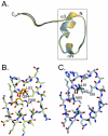
The macroH2A docking domain is unaffected by sequence changes. (A) Superposition of the docking domains (amino acid residues 80 through 119) of macroH2A (light gray) and Xla-H2A (light yellow) demonstrates that there are no differences in the path of the main chain between the two docking domains. (B and C) Detailed view of the boxed area (amino acids 80 through 105 in majorH2A) in panel A highlights significant amino acid differences (L83 to I80, Q84 to L81, and in particular, R88 to A85) that may alter the stability of this area. The sequence differences are shown in dark gray (macroH2A) and dark yellow (Xla-H2A). Also shown are some of the hydrogen bonds formed by R88 to stabilize this domain in Xla-H2A.
FIG. 4.
FIG. 4.
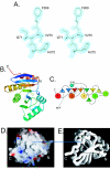
Structure of the nonhistone region of macroH2A. (A) Stereo view of a section of the  2Fo-Fc  electron density map, calculated at 1.6 Å and contoured at 2 σ, clearly showing a part of the sequence of the nonhistone region (F269-H272). (B) Overall structure of the nonhistone region (aa 180 through 370). The N terminus (N) is in blue and the C terminus (C) in red with a gradient of the colors of the visible spectrum in between. (C) A schematic representation of the fold in Fig. 1B. Beta-strands are depicted as arrowheads and helices as circles. (D) The surface representation of the nonhistone region. Basic regions are in blue, acidic regions in red, and neutral regions in white. (E) The C-α trace of the nonhistone region of macroH2A in exactly the same orientation as in panel D. The boxed area encompasses a large hydrophobic region that includes residues F183 to L186 and I356 to V360.
FIG. 5.
FIG. 5.
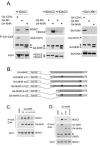
The nonhistone region (NHR) of macroH2A1.2 is associated with HDAC1. (A) Cos cells were cotransfected with 3 μg of expression vectors for Flag-tagged HDAC1, -2, or -3 or myc-tagged SUV39h1, and 3 μg of vectors expressing the NHR of macroH2A (aa 160 through 370) were fused to the DNA-binding domain of Gal4 (G4-NHR). The Gal4 binding domain alone (G4.BD) and G4-Cdyl were used as negative and positive controls, respectively. The Gal4 fusion proteins were immunoprecipitated from whole-cell extracts with 1 μg of an anti-Gal4 antibody, and proteins present in the complexes were analyzed with anti-Flag (recognizing HDAC1, HDAC2, and HDAC3) and anti-myc (recognizing SUV39h1) antibodies (upper panel). The same blot was then probed with an anti-Gal4 antibody (middle panel). G4.DB is not visible in the range of molecular weight shown (Fig. 4D). Asterisks indicate the Ig-heavy chains. The lower panels show the amounts of HDACs or SUV39h1 present in 5% of each input. (B) Parts of the NHR involved in the interaction with HDAC1. Schematic representations of G4-NHR constructs m1 and m2 have been generated by replacing 2 amino acids of hydrophobic patches (F182 and T183 in m1 and I360 and Y361 in m2) with arginines. The double mutant (m1/2) corresponds to the association of m1 and m2 mutations. (C and D) Coimmunoprecipitations of Flag-HDAC1 with G4-NHR proteins were performed as described in the legend to Fig. 1A, except that lysis of cells and washing of immunoprecipitated complexes were performed under more stringent conditions. Asterisks indicate the Ig-light chains. Wt, wild type.
FIG. 6.
FIG. 6.
The nonhistone region (NHR) of macroH2A1.2 is associated with hypoacetylated chromatin. (A) A total of 0.5 μg of each G4-NHR construct [or Gal4.DB or an empty vector (−) as controls] was cotransfected in Cos cells with 1 μg of a luciferase plasmid reporter containing five Gal4 sites in its promoter. In each transfection, 100 ng of pCMV-β-gal control plasmid was also used for normalization purposes. Luciferase activity was measured on cell extracts 24 h after transfection and normalized to that of β-galactosidase. Mean values of at least four independent assays are represented. Wt, wild type. (B) Schematic representation of the chromatin immunoprecipitation assay. Cos cells tranfected with expression vectors of either Ha-tagged H2A or Ha-tagged macroH2A in the presence of the ectopically expressed HAT p300 (in order to enhance background H3 acetylation to visualize HDAC activity) were lysed, and chromatin was fragmented by sonication in order to obtain DNA fragments with a mean size of 600 to 1,000 bp. Chromatin fragments containing nucleosomes with Ha-H2A or Ha-macroH2A were immunoprecipitated with an anti-Ha antibody and analyzed by silver staining or Western blotting for the presence of histones and the level of their acetylation. (C) Immunoprecipitated Ha-macroH2A and Ha-H2A were detected by anti-Ha Western blotting (lower right panel) and were also seen on a silver-stained gel (left panel, indicated with arrows). Acetylation level of coprecipitated histones was analyzed with antibodies recognizing acetylated histone H3 (third panel on the right) and compared to 5% of the input (upper right panel). Asterisks indicate the Ig light chains. The same blot was also probed with anti-H2A and anti-H3 antibodies (as indicated). (D and E) Nuclei of murine erythroleukemia cells were lysed and sonicated in order to generate small soluble chromatin fragments. (D) DNA analysis on an agarose electrophoresis gel showing that the fragments have a mean size of about 400 bp. (E) HDAC1, -2, or -3 was immunoprecipitated from the extracts using the specific respective antibodies (HDAC1, -2, or -3 IP), and the presence of macroH2A in the complex was detected by an anti-macroH2A Western blot (upper panel). The HDAC1 and -2 panels show the corresponding immunoprecipitated proteins as controls. Input corresponds to 5% of the extract. Asterisks indicate the Ig-heavy chains.

References

    1. Abbott, D. W., V. S. Ivanova, X. Wang, W. M. Bonner, and J. Ausio. 2001. Characterization of the stability and folding of H2A.Z chromatin particles: implications for transcriptional activation. J. Biol. Chem. 276:41945-41949. - PubMed
    1. Ahmad, K., and S. Henikoff. 2002. The histone variant H3.3 marks active chromatin by replication- independent nucleosome assembly. Mol. Cell 9:1191-1200. - PubMed
    1. Allen, M. D., A. M. Buckle, S. C. Cordell, J. Lowe, and M. Bycroft. 2003. The crystal structure of AF1521 a protein from Archaeoglobus fulgidus with homology to the non-histone domain of macroH2A. J. Mol. Biol. 330:503-511. - PubMed
    1. Angelov, D., A. Molla, P. Y. Perche, F. Hans, J. Cote, S. Khochbin, P. Bouvet, and S. Dimitrov. 2003. The histone variant MacroH2A interferes with transcription factor binding and SWI/SNF nucleosome remodeling. Mol. Cell 11:1033-1041. - PubMed
    1. Belotserkovskaya, R., S. Oh, V. A. Bondarenko, G. Orphanides, V. M. Studitsky, and D. Reinberg. 2003. FACT facilitates transcription-dependent nucleosome alteration. Science 301:1090-1093. - PubMed

Publication types

LinkOut - more resources