. 2005 Sep;25(17):7657-64.
doi: 10.1128/MCB.25.17.7657-7664.2005.

Reduction of spermatogenesis but not fertility in Creb3l4-deficient mice

Affiliations

Reduction of spermatogenesis but not fertility in Creb3l4-deficient mice

Ibrahim M Adham et al. Mol Cell Biol. 2005 Sep.

Abstract

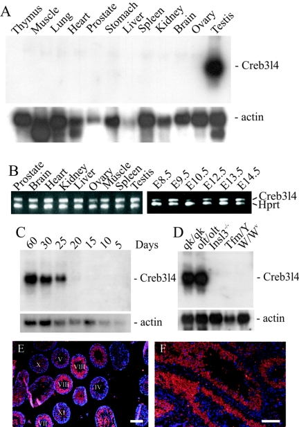
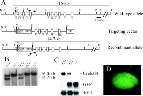
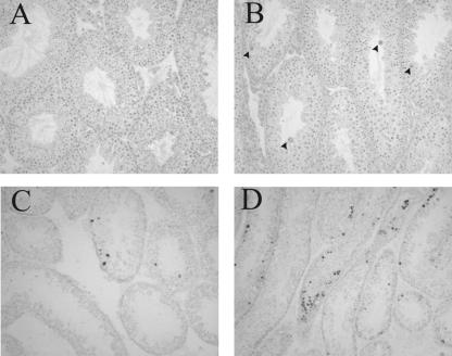
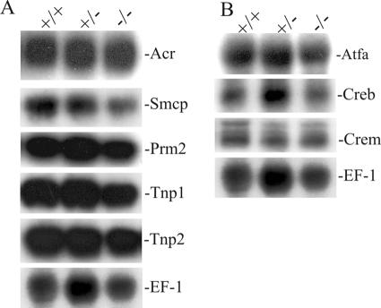
Creb3l4 belongs to the CREB/ATF family of transcription factors that are involved in mediating transcription in response to intracellular signaling. This study shows that Creb3l4 is expressed at low levels in all organs and in different stages of embryogenesis but is present at very high levels in the testis, particularly in postmeiotic male germ cells. In contrast to CREB3L4 in the human prostate, of which specific expression was detected, Creb3l4 transcripts in the mouse prostate could be detected only by RT-PCR. To identify the physiological function of Creb3l4, the murine gene was inactivated by replacement with the gene encoding green fluorescent protein. Surprisingly, Creb3l4-deficient mice were born at expected ratios, were healthy, and displayed normal long-term survival rates. Despite a significant reduction in the number of spermatozoa in the epididymis of Creb3l4(-)(/)(-) mice, the breeding of mutant males with wild-type females was productive and the average litter size was not significantly altered in comparison to wild-type littermates. Further analyses revealed that the seminiferous tubules of Creb3l4(-)(/)(-) mice contained all of the developmental stages, though there was evidence for increased apoptosis of meiotic/postmeiotic germ cells. These results suggest that Creb3l4 plays a role in male germ cell development, but its loss is insufficient to completely compromise the production of spermatozoa.


Figures

FIG. 1.
FIG. 1.
Expression analyses of the Creb3l4 gene. Northern blots with total RNA from different tissues of adult nice (A), from testis of 5-, 10-, 15, 20-, 25-, and 60-day-old mice (C) and from testis of different mutant mice (D) were hybridized with the Creb3l4 cDNA probe. Rehybridization with the β-actin cDNA revealed the level of RNA loading. (B) RT-PCR analysis with total RNA and Creb3l4-specific primers showed the presence of a 420-bp amplified fragment at all examined tissues and all embryonic stages. Production of the control Hprt products was observed throughout, demonstrating the presence of intact loaded RNA. (E and F) In situ hybridization of DIG-labeled Creb3l4 antisense transcripts to testis sections. The labeled antisense RNA was detected by streptavidin-Cy3 after signal amplification, and DNA was counterstained with DAPI (4′,6′-diamidino-2-phenylindole). The merged image shows the nuclei (blue) and the cytoplasmic localization of Creb3l4 transcripts (red). Bars, 100 μm.
FIG. 2.
FIG. 2.
Targeted disruption of the Creb3l4 gene. (A) Structure of the wild-type, targeted vector, and recombinant allele are shown together with the relevant restriction sites. The numbers under the rectangles indicate the 11 exons of Creb3l4. Predicted translation start sites (ATG) in the Creb3l4α and Creb3l4β isoforms are indicated by single stars. The Creb3l4α isoform is transcribed from exons 1a and 2 to 10, and its predicted translation start site is the first ATG. The Creb3l4β isoform contains the sequence of exons 1b and 2 to 10, and its open reading frame starts at the second ATG. A 4.0-kb XhoI/EcoRI fragment containing exon 1a to exon 4 was replaced by a GFP gene and a pgk-neo selection cassette (NEO). The 3′ external probe used and the predicted length of HindIII restriction fragments in Southern blot analysis are shown. The primers 1, 2, 3, and 4 used to amplify the wild-type and mutant allele by PCR are also indicated. Abbreviations: TK, thymidine kinase cassette; E, EcoRI; H, HindIII; N, NotI; P, PstI; X, XhoI. (B) Southern blot analysis of the recombinant ES cell clones. Genomic DNA extracted from ES cell clones was digested with HindIII and probed with the 3′ probe shown in panel A. The Creb3l4 wild-type allele generated a 16.0-kb HindIII fragment, whereas the targeted allele yielded a 14.7-kb HindIII fragment, as indicated in panel A. (C) Northern blot analysis. Total RNA of Creb3l4/, Creb3l4+/ and Creb3l4+/+ was hybridized with Creb3l4 and a GFP probe. Rehybridization of blots with human elongation factor-1 cDNA (EF-1) revealed the integrity of RNA loading. (D) GFP expression in the testis of a Creb3l4/ male. The GFP fluorescence is restricted to the seminiferous tubules.
FIG. 3.
FIG. 3.
Spermatogenesis in the testes of Creb3l4−/− mice. Histological sections from the testes of 12-week-old wild-type (A and C) and Creb3l4/ (B and D) mice are shown. (A and B) Staining with hematoxylin and eosin reveals the presence of all stages of spermatogenesis and the appearance of occasional multinucleated giant cells (arrows) in Creb3l4/ tubules. In situ TUNEL staining in the seminiferous tubules of wild-type (C) and Creb3l4/ (D) males revealed an increase in adluminal cells, with dark-stained nuclei in a subset of the tubules in mutant testes (D), while apoptotic cells in wild-type testes were most frequently located close to the basement membrane of seminiferous tubules (C). Magnification, ×200.
FIG. 4.
FIG. 4.
Analysis of gene expression in testes of Creb3l4−/− mice. Total RNA was isolated from testes of Creb3l4−/−, Creb3l4+/−, and Creb3l4+/+ mice, and 15 μg was analyzed by Northern blotting and sequential hybridization with radioactively labeled cDNA probes for the genes indicated. (A) Expression of germ cell-specific genes, which contain CREs in their promoters. (B) Expression of ATF/CREB genes. Rehybridization of blots with human elongation factor-1 cDNA (EF-1) revealed the integrity of RNA loading.

Similar articles

Cited by

References

    1. Aho, H., M. Schwemmer, D. Tessman, D. Murphy, G. Mattei, W. Engel, and I. M. Adham. 1996. Isolation, expression, and chromosomal localization of the human mitochondrial capsule selenoprotein gene (MCSP). Genomics 32:184-190. - PubMed
    1. Blendy, J. A., K. H. Kaestner, G. F. Weinbauer, E. Nieschlag, and G. Schutz. 1996. Severe impairment of spermatogenesis in mice lacking the CREM gene. Nature 380:162-165. - PubMed
    1. Cohen, P. E., O. Chisholm, R. J. Arceci, E. R. Stanley, and J. W. Pollard. 1996. Absence of colony-stimulating factor-1 in osteopetrotic (csfmop/csfmop) mice results in male fertility defects. Biol. Reprod. 55:310-317. - PubMed
    1. De Cesare, D., and P. Sassone-Corsi. 2000. Transcriptional regulation by cyclic AMP-responsive factors. Prog. Nucleic Acid Res. Mol. Biol. 64:343-369. - PubMed
    1. De Graeve, F., A. Bahr, B. Chatton, and C. Kedinger. 2000. A murine ATFa-associated factor with transcriptional repressing activity. Oncogene 19:1807-1819. - PubMed

Publication types

MeSH terms

Substances