. 2005 Sep;25(17):7854-67.
doi: 10.1128/MCB.25.17.7854-7867.2005.

Cationic sites on granzyme B contribute to cytotoxicity by promoting its uptake into target cells

Affiliations

Cationic sites on granzyme B contribute to cytotoxicity by promoting its uptake into target cells

Catherina H Bird et al. Mol Cell Biol. 2005 Sep.

Abstract

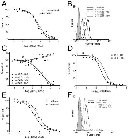
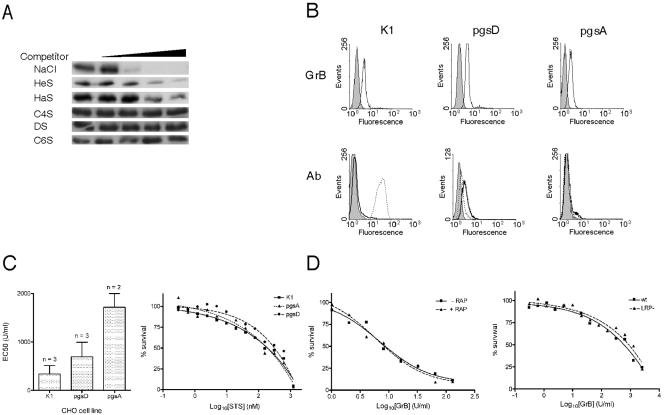
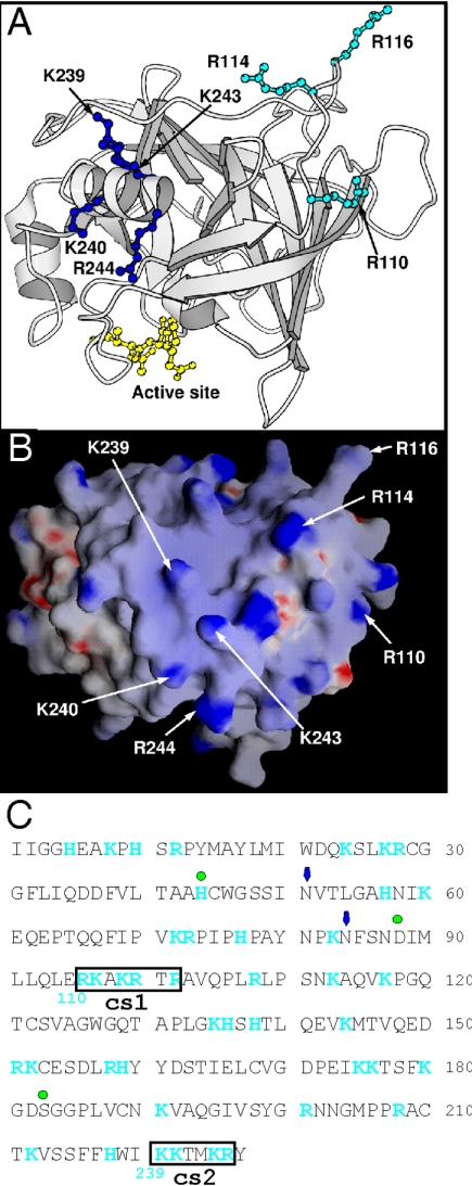
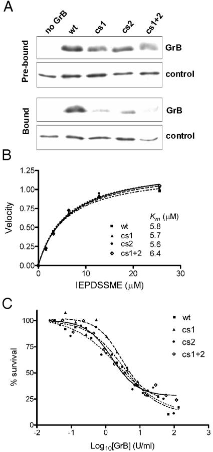
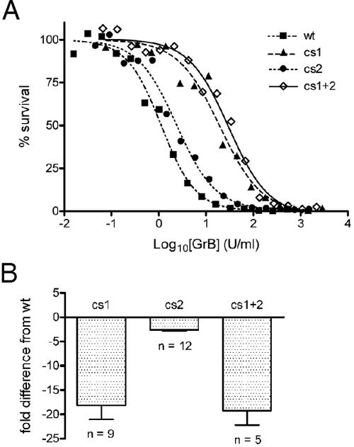
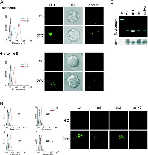
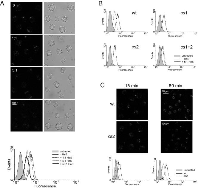
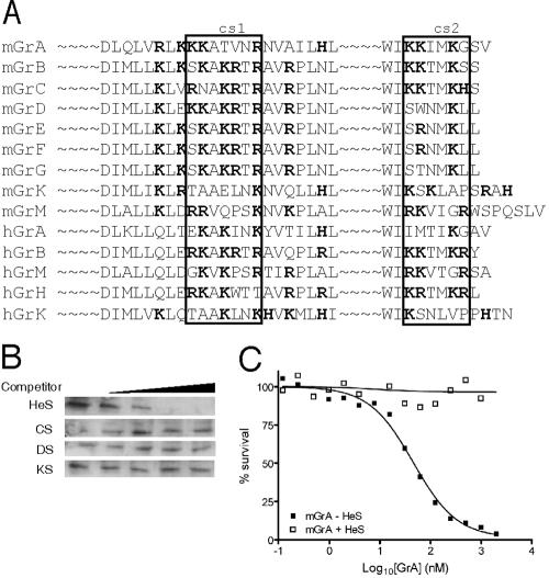
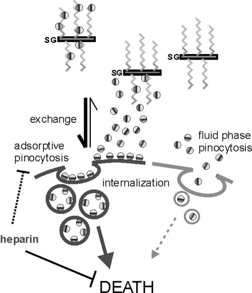
Granzyme B (GrB) is a key effector of cytotoxic lymphocyte-mediated cell death. It is delivered to target cells bound to the proteoglycan serglycin, but how it crosses the plasma membrane and accesses substrates in the cytoplasm is poorly understood. Here we identify two cationic sequences on GrB that facilitate its binding and uptake. Mutation of cationic sequence 1 (cs1) prevents accumulation of GrB in a distinctive intracellular compartment and reduces cytotoxicity 20-fold. Mutation of cs2 reduces accumulation in this intracellular compartment and cytotoxicity two- to threefold. We also show that GrB-mediated cytotoxicity is abrogated by heparin and that target cells deficient in cell surface sulfate or glycosaminoglycans resist GrB. However, heparin does not completely prevent GrB internalization and chondroitin 4-sulfate does not inhibit cytotoxicity, suggesting that glycosaminoglycans are not essential GrB receptors. We propose that GrB enters cells by nonselective adsorptive pinocytosis, exchanging from chondroitin sulfate on serglycin to anionic components of the cell surface. In this electrostatic "exchange-adsorption" model, cs1 and cs2 participate in binding of GrB to the cell surface, thereby promoting its uptake and eventual release into the cytoplasm.


Figures

FIG. 1.
FIG. 1.
Influence of man 6-P and electrostatic interactions on GrB uptake and cytotoxicity. (A) Native GrB and recombinant GrB kill target cells equally well. Enzymatic activities of the GrB preparations were matched by IETD-pNA cleavage assay. In the in vitro killing assay, Jurkat cells were incubated for 1 h at 37°C with a constant amount of perforin (empirically determined to lyse 10 to 15% of the cells) and increasing concentrations of GrB. Cells were washed and returned to complete medium, and survival was assessed 16 h later by MTT assay. The EC50 for recombinant GrB was 4.8 U/ml; for native GrB it was 5.4 U/ml. (B) Soluble man 6-P reduces uptake of native GrB but not recombinant GrB by Jurkat cells. Cells were incubated in 100 nM of FITC-GrB (recombinant) or 50 nM Alexa 488-GrB (native) in the absence or presence of 100 μM man 6-P at 37°C for 1 h and then washed and analyzed by FACS. (C) Heparin abrogates GrB-mediated cytotoxicity. GrB was preincubated at 37°C for 30 min with a 50-fold molar excess of heparin and then used in the in vitro killing assay. Heparin was maintained at at least a fivefold excess over GrB levels during exposure to cells. (D) Chondroitin 4-sulfate (CS) does not abrogate GrB-mediated cytotoxicity. Recombinant GrB was preincubated at 37°C for 30 min with a 50-fold molar excess of CS and then used in the in vitro killing assay. CS was maintained at at least a fivefold excess over GrB levels during exposure to cells. (E) Sodium chlorate treatment reduces killing of Jurkat cells by recombinant GrB. Target cells were cultured in the presence of 75 mM sodium chlorate for 48 h prior to and during the in vitro killing assay. The EC50 without chlorate was 3.4 U/ml; with chlorate it was 8.2 U/ml. (F) Chlorate treatment reduces uptake of FITC-GrB (recombinant) by Jurkat cells. Cells were cultured in the presence of 75 mM sodium chlorate for 48 h prior to GrB binding for 1 h at 37°C, washing, and FACS.
FIG. 2.
FIG. 2.
Contribution of GAGs and LRP to GrB-mediated cytotoxicity. (A) GrB binds GAGs. Recombinant GrB (50 nM) was added to 0, 0.1, 0.2, 0.3, or 0.4 M NaCl or to 0, 0.1, 1, 10, or 100 μM of the indicated GAG competitor and then incubated with heparin-Sepharose beads and washed. Bound GrB was removed from the beads by boiling in SDS and assessed by SDS-PAGE and immunoblotting. HeS, heparin; HaS, heparan sulfate; C4S, chondroitin 4-sulfate; DS, dermatan sulfate; C6S, chondroitin 6-sulfate. (B) GrB uptake does not require GAGs. Upper panels: CHO GAG-deficient cell lines were incubated with 600 nM FITC-GrB (recombinant) for 1 h at 37°C, washed, and analyzed by FACS. Lower panels: FACS analysis of cell surface GAGs on the CHO cells by binding of anti-HaS antibody (dotted line) or anti-CS antibody (solid line). (C) GAG-deficient CHO lines are less sensitive than wt to GrB but are equally sensitive to staurosporine (STS). Shown are the EC50s derived from repeated GrB in vitro killing assays (left panel) and an STS dose-response experiment as assessed by MTT assay (right panel). Error bars indicate the standard deviation. (D) In vitro killing of Jurkat cells by GrB is not inhibited by the LRP chaperone, RAP (left panel). LRP-deficient CHO cells remain sensitive to GrB in the in vitro killing assay (right panel).
FIG. 3.
FIG. 3.
Location of two clusters of positively charged residues on human GrB. The structure of human GrB bound to a peptide inhibitor has been solved (39) (Protein Database accession number: 1IAU). (A) The residues mutated to alanine in the GrB cs mutants are labeled (numbering in 1IAU is based by convention on chymotrypsin). R110, R114, and R116 (cs1) are in cyan ball-and-stick, and K239, K240, K243, and K244 (cs2) are in blue ball-and-stick. The peptide inhibitor Ac-IEPD-CHO is in yellow ball-and-stick, and indicates the position of the catalytic site. (B) GRASP electrostatic potential surface of GrB in an orientation similar to the image shown in panel A. The positively charged residues selected for mutagenesis are labeled. (C) Sequence of mature GrB showing positively charged residues (blue) and the cationic sequences (boxed). Green dots mark the residues of the catalytic triad, and arrows mark the positions of Asn-linked carbohydrate side chains. Numbering in blue below the sequence is based on chymotrypsin.
FIG. 4.
FIG. 4.
GrB cs mutants have impaired GAG binding but normal catalytic and apoptotic function. (A) Heparin-binding of cs mutants. Equal amounts of wt or mutant GrB were mixed with heparin-Sepharose beads. An irrelevant heparin-binding control protein (antithrombin) was included as a tracer to monitor loss of beads during the procedure. Samples were assessed by SDS-PAGE and immunoblotting. The Prebound panel compares the input GrBs used in the experiment, with no binding or washing steps, as assessed immediately after mixing with the tracer. The Bound panel shows the GrB remaining after binding and washing. Results are representative of five independent experiments. (B) The catalytic activity of wt and cs mutants on a quenched-fluorescence substrate Abz-IEPDSSMESK-dnp was measured as described previously (46). (C) The apoptotic function of the cs mutants was assessed using a modified in vitro killing assay, in which perforin was replaced by SLO. Enzymatic activities of the granzymes were matched via IETD-pNA cleavage assay. Jurkat cells were incubated for 1 h at 37°C with an empirically determined amount of SLO (permeabilizes 80% of cells) and increasing concentrations of GrB. Cells were washed, and survival was assessed 16 h later by MTT assay. The EC50 for wt GrB was 5 U/ml.
FIG. 5.
FIG. 5.
GrB cs mutants show impaired cytotoxicity in the presence of perforin. (A) Representative in vitro killing assay comparing wt and mutant GrBs. See Fig. 1A legend for details. (B) Severalfold differences in EC50s between the cs mutants and wt GrB in the in vitro killing assays. Error bars indicate the standard deviation. The EC50 for wt GrB was 3.9 ± 1.8 U/ml (n = 19).
FIG. 6.
FIG. 6.
Uptake and intracellular localization of GrB and the cs mutants. Binding and accumulation of FITC-labeled transferrin (125 nM) or FITC-labeled granzymes (600 nM) in the presence of sublytic perforin. Jurkat cells were exposed to fluoresceinated protein and perforin at 4°C or 37°C for 15 min, washed, and analyzed by FACS. Autofluorescence levels of cells treated with perforin alone at 4°C (shaded histogram) or 37°C (black line) were identical. Cells from the same populations were also analyzed by CLSM. (A) Uptake and localization of wt GrB compared to transferrin. Image shows the distribution of fluoresceinated protein (FITC) with a corresponding differential interference contrast image. Shown are single optical sections from single cells (left and center panels) or z-stacks of multiple cells (right panels). (B) Uptake and localization of cs mutants at 4°C and 37°C was analyzed as described above. Images show single optical sections. (C) A fluorograph and an immunoblot probed with anti-GrB monoclonal 2C5 demonstrate integrity and equivalent FITC labeling of wt GrB and the cs mutants. The fluorograph was taken prior to transfer to a membrane for immunoblotting (Tf, transferrin).
FIG. 7.
FIG. 7.
Effect of heparin on uptake and localization of granzymes, and the role of cs2. Jurkat cells were exposed to perforin and FITC-GrB at 37°C for 15 min or 60 min (C), washed, and analyzed by FACS and/or CLSM. (A) Heparin reduces but does not prevent GrB accumulation in intracellular vesicles. Images show z-stacks of multiple cells: the corresponding FACs profile is shown beneath. (B) A fivefold excess of heparin reduces GrB accumulation in Jurkat cells but does not further reduce accumulation of cs mutants. (C) cs2 shows reduced uptake and vesicular accumulation. Shown are z stacks of multiple cells.
FIG. 8.
FIG. 8.
Electrostatic interactions may contribute to the function of other granzymes. (A) Regions homologous to cs1 and cs2 (boxed) in other human and mouse granzymes. Positively charged amino acids are indicated in bold. (B) GrA binds GAGs. A 50 nM concentration of recombinant mouse GrA was used as described in the legend to Fig. 2A. (C) Heparin abrogates GrA and perforin-mediated killing of Jurkat cells. Recombinant GrA was preincubated at 37°C for 30 min with a 50-fold molar excess of heparin and then used in the in vitro killing assay (see Fig. 1A for details.) Heparin was maintained at at least a fivefold excess over GrA levels during exposure to cells.
FIG. 9.
FIG. 9.
The exchange/adsorption model. Granzyme (indicated by two-tone sphere) is delivered into the synapse electrostatically bound to the CS chains (zigzag lines) of serglycin (SG). Granzyme exchanges from CS chains to more negatively charged components of the target cell surface and is internalized by adsorptive pinocytosis (7). Mutation of cs1 or cs2 compromises the cationic surface (indicated by darker hemisphere) of granzyme B and reduces adsorption. Nonadsorbed wt or mutant granzyme is internalized by fluid-phase pinocytosis. Perforin (not shown) releases granzyme from either pathway to cause cell death. Pretreatment of granzyme with excess heparin substantially reduces adsorption but not fluid-phase uptake and prevents death by interfering with an additional unidentified step(s) common to both pathways. Aspects of this model are consistent with recent studies showing that SG-GrB complexes are not internalized (37) and inhibition of granzyme uptake by anionic competitors (41).

References

    1. Baeuerle, P. A., and W. B. Huttner. 1986. Chlorate—a potent inhibitor of protein sulfation in intact cells. Biochem. Biophys. Res. Commun. 141:870-877. - PubMed
    1. Barry, M., and C. R. Bleackley. 2002. Cytotoxic lymphocytes: all roads lead to death. Nature Rev. Immunol. 2:401-409. - PubMed
    1. Bretthauer, R. K., and F. J. Castellino. 1997. Glycosylation of Pichia pastoris-derived proteins. Biotechnol. Appl. Biochem. 30:193-200. - PubMed
    1. Browne, K. A., E. Blink, V. R. Sutton, C. J. Froelich, D. A. Jans, and J. A. Trapani. 1999. Cytosolic delivery of granzyme B by bacterial toxins: evidence that endosomal disruption, in addition to transmembrane pore formation, is an important function of perforin. Mol. Cell. Biol. 19:8604-8615. - PMC - PubMed
    1. Cardin, A. D., and H. J. Weintraub. 1989. Molecular modeling of protein-glycosaminoglycan interactions. Arteriosclerosis 9:21-32. - PubMed

Publication types

MeSH terms

LinkOut - more resources