. 2005 Sep;25(17):7868-78.
doi: 10.1128/MCB.25.17.7868-7878.2005.

c-Myc is required for the formation of intestinal crypts but dispensable for homeostasis of the adult intestinal epithelium

Affiliations

c-Myc is required for the formation of intestinal crypts but dispensable for homeostasis of the adult intestinal epithelium

Michael D Bettess et al. Mol Cell Biol. 2005 Sep.

Abstract

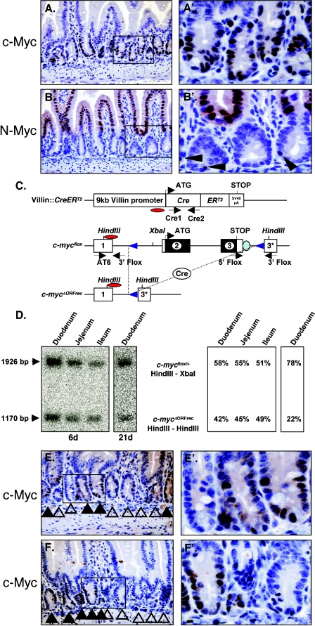
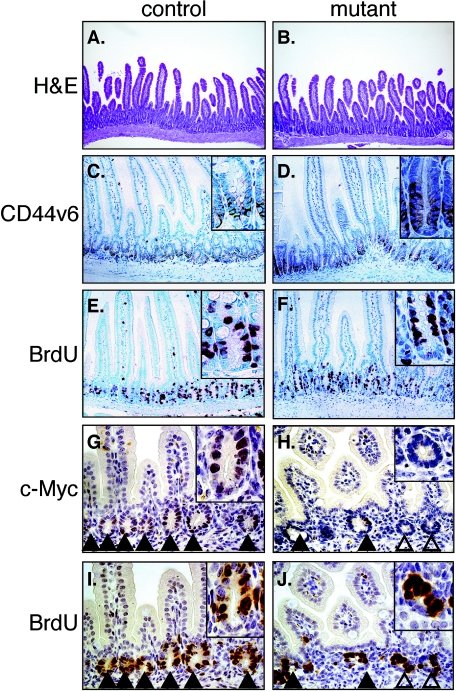
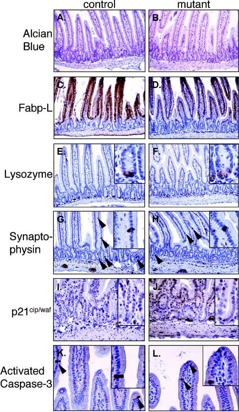
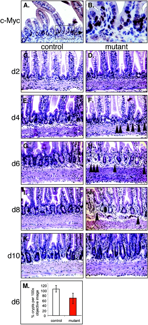
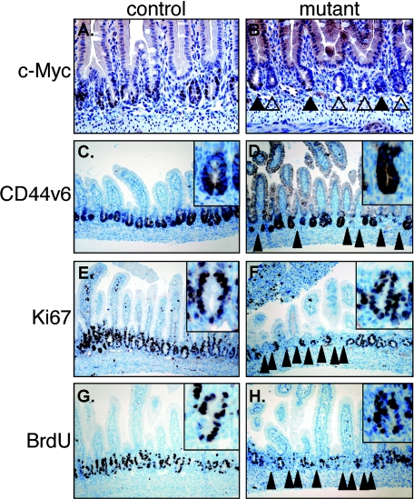
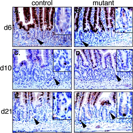
In self-renewing tissues such as the skin epidermis and the bone marrow, Myc proteins control differentiation of stem cells and proliferation of progenitor cell types. In the epithelium of the small intestine, we show that c-Myc and N-Myc are expressed in a differential manner. Whereas c-Myc is expressed in the proliferating transient-amplifying compartment of the crypts, N-Myc is restricted to the differentiated villus epithelium and a single cell located near the crypt base. c-Myc has been implicated as a critical target of the canonical Wnt pathway, which is essential for formation and maintenance of the intestinal mucosa. To genetically assess the role of c-Myc during development and homeostasis of the mammalian intestine we induced deletion of the c-myc(flox) allele in the villi and intestinal stem cell-bearing crypts of juvenile and adult mice, via tamoxifen-induced activation of the CreER(T2) recombinase, driven by the villin promoter. Absence of c-Myc activity in the juvenile mucosa at the onset of crypt morphogenesis leads to a failure to form normal numbers of crypts in the small intestine. However, all mice recover from this insult to form and maintain a normal epithelium in the absence of c-Myc activity and without apparent compensation by N-Myc or L-Myc. This study provides genetic and molecular evidence that proliferation and expansion of progenitors necessary to maintain the adult intestinal epithelium can unexpectedly occur in a Myc-independent manner.


Figures

FIG.1.
FIG.1.
Myc expression and villin::CreERT2-mediated recombination of the c-mycflox allele in the small intestine. (A) Immunohistochemical analysis of c-Myc in the adult duodenum. Enlargement of the indicated box is shown in A′. (B and B′) Immunohistochemistry of N-Myc in adult duodenum. (C) Schematic representation of the villin::CreERT2 and c-mycflox alleles used. The c-mycΔORFrec allele represents the locus after Cre mediated recombination. The three c-myc exons are illustrated as boxes; regions in black indicate the open reading frame. Restriction sites are shown on top. Red ovals indicate probe used for Southern blot analysis. Arrows below each scheme indicate locations of the primers used for genotyping. (D) Southern blot analysis of villin::CreERT2; c-mycflox/+ mice in the small intestine 6 days (duodenum, ileum, and jejunum) and 21 days (duodenum) after the first of four consecutive tamoxifen injections. The c-myc probe hybridizes to the floxed and wild-type alleles (1,926 bp) and to the deleted c-mycΔORFrec allele (ΔORFrec: 1,170 bp) in recombined cells. (E and F) Immunohistochemical analysis of c-Myc in duodenal tissue sections derived from villin::CreERT2, c-mycΔORF/flox mice 21 days following the first injection of tamoxifen. Solid arrows indicate c-Myc-expressing crypts (c-mycΔORF/flox). Open arrows indicate c-Myc-negative crypts (c-mycΔORF/ORFrec).
FIG. 2.
FIG. 2.
Histological analysis and marker gene expression in small intestines of control (A, C, E, G, and I) and mutant (B, D, F, H, and J) mice. (A and B) Hematoxylin/eosin staining. Magnification, 100×. (C and D) Immunohistochemical analysis of CD44v6 expression. (E and F) Bromodeoxyuridine (BrdU) incorporation. (G to J) Immunohistochemical analysis of serial sections of the duodenum detecting c-Myc expression (G and H) and bromodeoxyuridine incorporation (I and J). Solid arrows indicate c-Myc-expressing crypts (c-mycΔORFrec/flox) and open ones indicate c-Myc-negative crypts (c-mycΔORFrec/ΔORFrec). (A to F) Adult mice (G to J) P14 juvenile mice. Magnification, 200×.
FIG. 3.
FIG. 3.
Expression analysis of differentiation and apoptosis markers in control (A, C, E, G, I, and K) and mutant (B, D, F, H, J, and L) duodenum. (A and B) Alcian Blue staining to detect goblet cells. (C and D) Fabp-L expression to detect enterocytes. (E and F) Lysozyme expression in Paneth cells. (G and H) Synaptophysin expression in endocrine cells. (I and H) Expression of the CDK inhibitor p21cip/waf. (K and L) expression of activated caspase-3 to monitor apoptotic cells. Arrows indicate lysozyme, synaptophysin or activated caspase-3-positive epithelial cells.
FIG. 4.
FIG. 4.
Kinetic analysis of the crypt formation process in normal (A, B, C, E, G, I, and K) and mutant (D, F, H, J, and L) mice. (A and B) c-Myc expression in the intervillus region of seven-day-old mice. (C to L) Seven-day-old control or mutant mice were injected with tamoxifen and the duodenum was analyzed by hematoxylin/eosin staining every second day for 10 days. (M) Graphical representation of the percentage of crypts per phase contrast image (100× objective) of tissue sections derived from six different littermates of control (100%) and mutant mice (red bar) 6 days after the first tamoxifen injection. Solid arrows in F, H, and J indicate areas in which crypt formation has not occurred.
FIG. 5.
FIG. 5.
Immunohistochemical analysis of c-Myc and crypt markers in juvenile duodenal tissue sections of control (A, C, E, and G) and mutant (B, D, F, and H) mice 6 days after Cre induction. (A and B) c-Myc; (C and D) CD44v6; (E and F) Ki67; (G and H) bromodeoxyuridine (BrdU). Solid arrows indicate c-Myc-positive crypts. Open arrows indicate c-Myc negative crypts. Solid pointed arrowheads in panels D, F, and H indicate areas lacking normal crypt formation.
FIG. 6.
FIG. 6.
Immunohistochemical analysis of N-Myc expression in control (A, C, and E) or mutant (B, D, and F) duodenum after Cre induction at the time points indicated. Arrowheads indicate single N-Myc-expressing cells at the bottom of crypts.

Similar articles

Cited by

References

    1. Alarcon, R. M., B. A. Rupnow, T. G. Graeber, S. J. Knox, and A. J. Giaccia. 1996. Modulation of c-Myc activity and apoptosis in vivo. Cancer Res. 56:4315-4319. - PubMed
    1. Amati, B., K. Alevizopoulos, and J. Vlach. 1998. Myc and the cell cycle. Front Biosci. 3:D250-68. - PubMed
    1. Anderson, C. B., K. L. Neufeld, and R. L. White. 2002. Subcellular distribution of Wnt pathway proteins in normal and neoplastic colon. Proc. Natl. Acad. Sci. USA 99:8683-8688. - PMC - PubMed
    1. Askew, D. S., R. A. Ashmun, B. C. Simmons, and J. L. Cleveland. 1991. Constitutive c-myc expression in an IL-3-dependent myeloid cell line suppresses cell cycle arrest and accelerates apoptosis. Oncogene 6:1915-1922. - PubMed
    1. Augenlicht, L. H., S. Wadler, G. Corner, C. Richards, L. Ryan, A. S. Multani, S. Pathak, A. Benson, D. Haller, and B. G. Heerdt. 1997. Low-level c-myc amplification in human colonic carcinoma cell lines and tumors: a frequent, p53-independent mutation associated with improved outcome in a randomized multi-institutional trial. Cancer Res. 57:1769-1775. - PubMed

Publication types

Substances

LinkOut - more resources