. 2005 Aug 18;436(7053):1020-1024.
doi: 10.1038/nature03885.

Local translation of RhoA regulates growth cone collapse

Affiliations

Local translation of RhoA regulates growth cone collapse

Karen Y Wu et al. Nature. .

Abstract

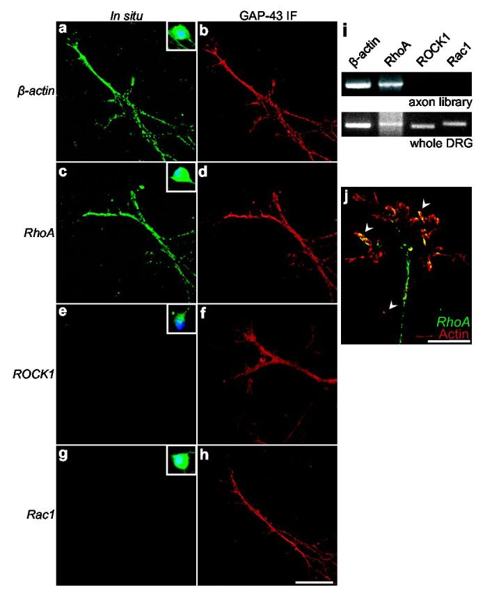
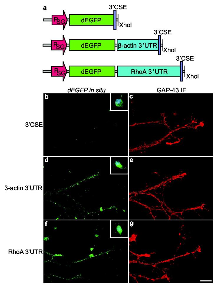
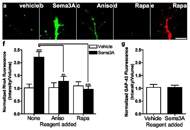
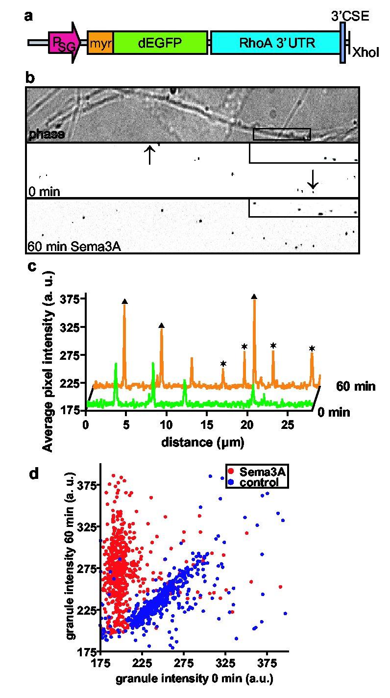
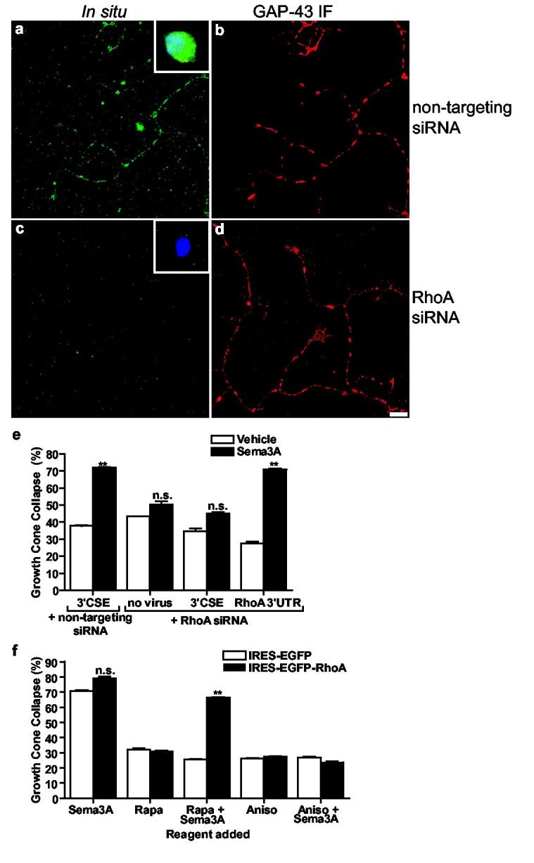
Neuronal development requires highly coordinated regulation of the cytoskeleton within the developing axon. This dynamic regulation manifests itself in axonal branching, turning and pathfinding, presynaptic differentiation, and growth cone collapse and extension. Semaphorin 3A (Sema3A), a secreted guidance cue that primarily functions to repel axons from inappropriate targets, induces cytoskeletal rearrangements that result in growth cone collapse. These effects require intra-axonal messenger RNA translation. Here we show that transcripts for RhoA, a small guanosine triphosphatase (GTPase) that regulates the actin cytoskeleton, are localized to developing axons and growth cones, and this localization is mediated by an axonal targeting element located in the RhoA 3' untranslated region (UTR). Sema3A induces intra-axonal translation of RhoA mRNA, and this local translation of RhoA is necessary and sufficient for Sema3A-mediated growth cone collapse. These studies indicate that local RhoA translation regulates the neuronal cytoskeleton and identify a new mechanism for the regulation of RhoA signalling.


Figures

Figure 1.
Figure 1.
RhoA mRNA is localized in axons and growth cones. In situ hybridization experiments using digoxigenin-labeled riboprobes directed against a, β-actin, c, RhoA, e, ROCK1, and g, Rac1 transcripts. β-actin and RhoA transcripts are localized to axons while Rac1 and ROCK1 transcripts are not detectable. Insets (a,c,e,g), In situ hybridization of cell bodies of dissociated DRG neurons. DAPI staining is shown in blue. b,d,f,h, GAP-43 immunofluorescence of axons in (a,c,e,g), respectively. Scale bar, 20 μm. i, Detection of RhoA mRNA in axons by RT-PCR from purified axons. Only transcripts for RhoA and β-actin were detected in axonal preparations (upper panel), while all transcripts were detected when whole explant RNA was used as a template (lower panel). j, Growth cones were visualized by actin immunofluorescence (red). Arrowheads indicate RhoA mRNA (green) along the periphery of the growth cone. Scale bar, 10 μm.
Figure 2.
Figure 2.
The RhoA 3’UTR contains an axonal targeting element. a, Schematic of Sindbis constructs. Transcripts encoding destabilized EGFP (dEGFP) contained either the conserved sequence element (3’CSE) of Sindbis as the 3’UTR of the transcript, the 3’UTR of RhoA, or the 3’UTR of β-actin. PSG, viral subgenomic promoter. b,d,f, In situ hybridization using riboprobes specific for dEGFP. dEGFP transcripts containing either the 3’UTR of β-actin or RhoA were targeted to axons. dEGFP transcripts containing only the viral 3’CSE sequence were not detected in axons. Insets (b,d,f), In situ hybridization of cell bodies. c,e,g, GAP-43 immunofluorescence of axons in (b,d,f).
Figure 3.
Figure 3.
Sema3A induces RhoA translation. Severed axons were preincubated with vehicle, 10 nM rapamycin, or 40 μM anisomycin, treated with Sema3A for 60 min, and RhoA was detected by immunofluorescence and normalized to axonal volume using GAP-43-defined cellular borders. a,b, Sema3A treatment increases RhoA levels. c, Sema3A-induced increases in RhoA are blocked by anisomycin. d, Sema3A-induced increases in RhoA are blocked by rapamycin. e, Example of GAP-43 immunofluorescence staining of the axon terminal seen in (d). Scale bar, 10 μm. f, Summary of results from a-d. n=50 growth cones per condition **p < 0.01. g, Normalized GAP-43 immunofluorescence is unaltered by Sema3A treatment. n=50 growth cones per condition.
Figure 4.
Figure 4.
Sema3A activates translation of a RhoA reporter. a, Schematic of Sindbis reporter construct. The reporter contains a myristoylated, destabilized EGFP (d1EGFP) with the 3’UTR of RhoA. b, Sema3A induces translation of the reporter. Phase (top) and EGFP fluorescence (middle) images of axons. Fluorescence images are shown with inverted contrast to facilitate visualization. Lower panel, Newly formed puncta and puncta with increased signal intensity are seen following Sema3A treatment. Inset, a region of the axon in the top panel is magnified to show changes in puncta number and intensity. Scale bar, 10 μm. c, A line scan performed at 0 (green) and 60 min (orange) in the region demarcated by arrows. Newly formed puncta (asterisks) and puncta with increased signal intensity (triangles) are seen following Sema3A treatment. d, Scatter plot of puncta intensities at 0 and 60 min. 580 puncta from experiments monitoring Sema3A- (red) and vehicle-treated (blue) axons were plotted. The majority of points from Sema3A-treated axons remained above the diagonal, indicating newly formed puncta or pre-existing puncta that increased in intensity upon Sema3A treatment. Most points from vehicle-treated axons remained along the diagonal, indicating that puncta were largely unaffected by vehicle treatment. The background levels in these images averaged approximately 185.
Figure 5.
Figure 5.
Axonal RhoA translation mediates Sema3A signaling. a-d, siRNA-mediated knockdown of RhoA transcripts in axons. In situ hybridization of RhoA transcripts in DRG axons was unaffected by control siRNA (a), and abolished in neurons transfected RhoA 5'UTR-directed siRNA (c). GAP-43 immunofluorescence (b,d) of axons in a,c. Insets, cell body staining. Scale bar, 10 μm. e, Axonal RhoA transcripts are required for Sema3A-induced collapse. Endogenous RhoA was knocked down with RhoA 5'UTR-directed siRNA, and RhoA was restored via Sindbis pseudoviruses expressing EGFP-RhoA3’CSE or EGFP-RhoA3’RhoA. The domain targeted by the siRNA is absent from the viral constructs. Sema3A-mediated collapse was significantly reduced in RhoA siRNA-transfected neurons infected with pseudovirus expressing EGFP-RhoA3’CSE, but was restored in RhoA siRNA-transfected neurons infected with pseudovirus expressing EGFP-RhoA3’RhoA. n=50 growth cones per condition, **p <0.01. f, Sema3A-mediated growth cone collapse was blocked by rapamycin (10 nM) in axons expressing IRES-EGFP and restored in axons expressing IRES-EGFP-RhoA. n=50 growth cones per condition, **p <0.01.

References

    1. Gallo G, Letourneau PC. Regulation of growth cone actin filaments by guidance cues. Journal of Neurobiology. 2004;58:92–102. - PubMed
    1. Campbell DS, Holt CE. Chemotropic responses of retinal growth cones mediated by rapid local protein synthesis and degradation. Neuron. 2001;32:1013–26. - PubMed
    1. Luo Y, Raible D, Raper JA. Collapsin: a protein in brain that induces the collapse and paralysis of neuronal growth cones. Cell. 1993;75:217–227. - PubMed
    1. Kolodkin AL, et al. Neuropilin is a semaphorin III receptor. Cell. 1997;90:753–762. - PubMed
    1. He Z, Tessier-Lavigne M. Neuropilin is a receptor for the axonal chemorepellent Semaphorin III. Cell. 1997;90:739–751. - PubMed

Publication types

MeSH terms