. 2005 Aug 18:5:17.
doi: 10.1186/1471-213X-5-17.

Inactivation of the Huntington's disease gene (Hdh) impairs anterior streak formation and early patterning of the mouse embryo

Affiliations

Inactivation of the Huntington's disease gene (Hdh) impairs anterior streak formation and early patterning of the mouse embryo

Juliana M Woda et al. BMC Dev Biol. .

Abstract

Background: Huntingtin, the HD gene encoded protein mutated by polyglutamine expansion in Huntington's disease, is required in extraembryonic tissues for proper gastrulation, implicating its activities in nutrition or patterning of the developing embryo. To test these possibilities, we have used whole mount in situ hybridization to examine embryonic patterning and morphogenesis in homozygous Hdh(ex4/5) huntingtin deficient embryos.

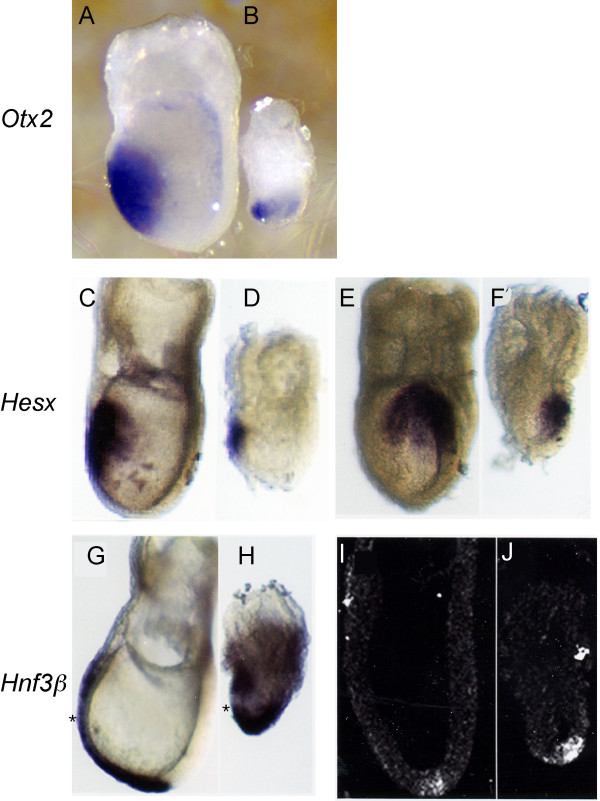
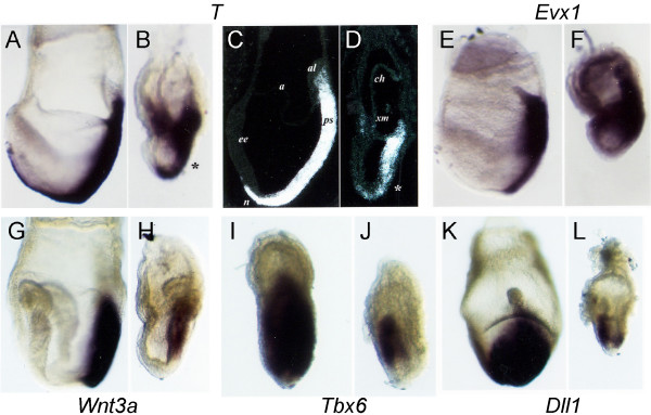
Results: In the absence of huntingtin, expression of nutritive genes appears normal but E7.0-7.5 embryos exhibit a unique combination of patterning defects. Notable are a shortened primitive streak, absence of a proper node and diminished production of anterior streak derivatives. Reduced Wnt3a, Tbx6 and Dll1 expression signify decreased paraxial mesoderm and reduced Otx2 expression and lack of headfolds denote a failure of head development. In addition, genes initially broadly expressed are not properly restricted to the posterior, as evidenced by the ectopic expression of Nodal, Fgf8 and Gsc in the epiblast and T (Brachyury) and Evx1 in proximal mesoderm derivatives. Despite impaired posterior restriction and anterior streak deficits, overall anterior/posterior polarity is established. A single primitive streak forms and marker expression shows that the anterior epiblast and anterior visceral endoderm (AVE) are specified.

Conclusion: Huntingtin is essential in the early patterning of the embryo for formation of the anterior region of the primitive streak, and for down-regulation of a subset of dynamic growth and transcription factor genes. These findings provide fundamental starting points for identifying the novel cellular and molecular activities of huntingtin in the extraembryonic tissues that govern normal anterior streak development. This knowledge may prove to be important for understanding the mechanism by which the dominant polyglutamine expansion in huntingtin determines the loss of neurons in Huntington's disease.


Figures

Figure 1
Figure 1
AVE displacement and anterior neurectoderm induction occur normally in the absence of huntingtin. Whole mount in situ hybridization analysis of Otx2 (A,B) and Hesx (C-F) in E7.5 normal (A,C,E) and mutant (B,D,F) embryos reveals that neuroectoderm and anterior visceral endoderm (AVE) develop normally in huntingtin deficient embryos, although the neuroectoderm expression domain is reduced. Asymmetrical expression of Hesx in mutant embryos (F) suggests that left-right transcriptional control is maintained. Hnf3β expression in the definitive endoderm extends around the distal tip and is reduced in the AVE (*) in both normal (G,I) and mutant embryos (H,J). Taken together, these results suggest normal ectoderm and endoderm induction and localization in Hdhex4/5/Hdhex4/5 embryos. Embryos are shown in lateral views, with anterior to the left in all pictures with the exception of E and F. Embryos are viewed from the anterior aspect in E and F.
Figure 2
Figure 2
Huntingtin is required for formation of anterior primitive streak and paraxial mesoderm. Whole mount and section insitu hybridizations of E7.5 embryos shows T (Brachyury) (A-D) is expressed in the primitive streak, node, axial mesoderm and Evx1 (E-F) is expressed in the primitive streak, most strongly in the proximal streak wild-type embryos. However, in mutant embryos, both T (B, D) and Evx1 (F) are ectopically expressed in the extraembryonic region. Wnt3A expression is reduced in mutant embryos (H), although the localization of its expression to the proximal streak is the same as in wild-type embryos (G). Analysis of paraxial mesoderm markers Tbx6 (I,J) and Dll1 (K,L), reveals that these markers are reduced in mutant embryos (J,L), suggesting impaired paraxial mesoderm production in the absence of huntingtin. Embryos in A-H are shown in a lateral view with anterior oriented to the left. Embryos in I-L are shown in a posterior view (I,K) or near posterior (J,L) view with proximal oriented toward the top. In (C,D), al = allantois, a = amnion, ch = chorion, ee = embryonic node(N), em = extraembryonic mesoderm, n = node, ps = primitive streak. Rather than a node, mutant embryos exhibit a region of disorganized cells (*) at the distal extent of the short primitive streak.
Figure 3
Figure 3
Impaired regional restriction of gene expression in huntingtin deficient embryos. X-gal staining of Nodal-LacZ embryos shows staining in endoderm near the node of normal embryos (A,C) but broad staining in mutant embryos (B, D), although expression is higher in the posterior. The tight node expression of Nodal in normal embryos (C) is lost in mutant embryos (D), consistent with the loss of a morphological node in the absence of huntingtin. Whole mount and in situ hybridization of E7.5 day embryos reveals that Fgf8 is detected in the proximal streak and is downregulated in cells migrating out of the streak in normal embryos (E,G). In contrast, Fgf8 remains highly expressed in mutant embryos (F,H). Transient expression of Gsc in the definitive endoderm overlying the prospective head region in normal embryos (I,K) is distinguished in other cell layers in normal embryos but remains unrestricted in mutant embryos (J,L). Earlier posterior expression of Gsc is also maintained in mutant embryos (J) while it is down-regulated in normal embryos (I). Embryos (A,B,E,F,G,H,I,K,L) are shown in a lateral view with anterior oriented to the left. Embryos (C,D) are in a posterior view.
Figure 4
Figure 4
Normal expression of extraembryonic markers in huntingtin deficient embryos. Whole mount in situ hybridization analysis at E7.5 of markers of the extraembryonic tissues reveals grossly normal expression in the absence of huntingtin. Hnf4, expressed in the visceral endoderm at the junction of embryonic-ectoderm junction (A), is normal in mutant embryos, although the signal is slightly higher (B). Similarly, the expression of Pem transcripts is maintained in mutant embryos (D) similar to normal embryos (C), although Pem is expressed in the abnormal lopsided overhang of visceral endoderm over the anterior of the mutant embryos. Expression of extraembryonic signaling molecules is unaffected by the loss of huntingtin, as evidenced by the expression of Bmp4 (E,F) in the extraembryonic ectoderm, and Lefty1 and Dkk1 (I-L) in the AVE in mutant embryos. Bmp4 is not localized, however, to a ring of extraembryonic ectoderm in mutant embryos (F) as in normal embryos (E). Primitive germ cells (PCGs) are induced normally in both wild-type (G) and mutant embryos (H), suggesting the Bmp4 signaling from the extraembryonic ectoderm to the epiblast is normal. Lefty1 expression appears disorganized in mutant embryos (I) compared to wild-type embryos (J). In contrast, the anterior expression of Dkk1 in the AVE in mutant embryos (L) matches the wild-type expression pattern (K). Despite normal AVE formation, head folds fail to form in mutant embryos, even when cultured in nutrient rich media for 24 hours. Wild-type E7.5 embryos, when cultured in 75% rat serum, develop somites (M), heart (white arrow, N) and head folds (blue arrow head, N) in culture. In contrast, huntingtin deficient embryos continue to live in culture but do not form headfolds, heart or somites (O). Embryos are shown in a lateral view (A-F, I-J) with anterior oriented to the left. Embryos in (G,H,K,L) are shown in an anterior view with proximal oriented up.

References

    1. Huntington's Disease Collaborative Research Group A novel gene containing a trinucleotide repeat that is expanded and unstable on Huntington's disease chromosomes. Cell. 1993;72:971–83. doi: 10.1016/0092-8674(93)90585-E. - DOI - PubMed
    1. Gusella JF, MacDonald ME. Molecular genetics: unmasking polyglutamine triggers in neurodegenerative disease. Nat Rev Neurosci. 2000;1:109–15. doi: 10.1038/35039051. - DOI - PubMed
    1. Takano H, Gusella JF. The predominantly HEAT-like motif structure of huntingtin and its association and coincident nuclear entry with dorsal, an NF-κB/REL/dorsal family transcription factor. BMC Neuroscience . 2002;3:15. doi: 10.1186/1471-2202-3-15. - DOI - PMC - PubMed
    1. Andrade MA, Bork P. HEAT repeats in the Huntington's disease protein. Nat Genet. 1995;11:115–6. doi: 10.1038/ng1095-115. - DOI - PubMed
    1. MacDonald ME. Huntingtin: alive and well and working in middle management. Sci STKE. 2003;2003:pe48. - PubMed

Publication types