Comparative Study
. 2005 Nov 1;568(Pt 3):815-40.
doi: 10.1113/jphysiol.2005.093948. Epub 2005 Aug 18.

Mechanisms of target-cell specific short-term plasticity at Schaffer collateral synapses onto interneurones versus pyramidal cells in juvenile rats

Affiliations
Comparative Study

Mechanisms of target-cell specific short-term plasticity at Schaffer collateral synapses onto interneurones versus pyramidal cells in juvenile rats

Hua Yu Sun et al. J Physiol. .

Abstract

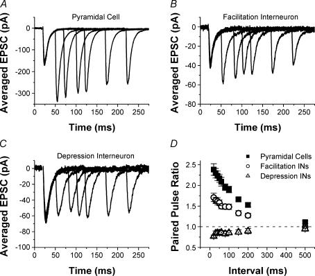
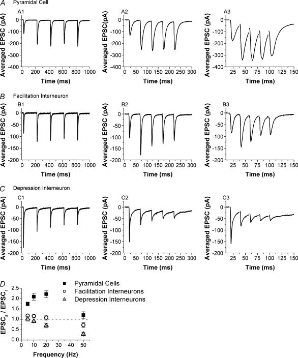
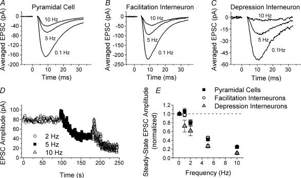
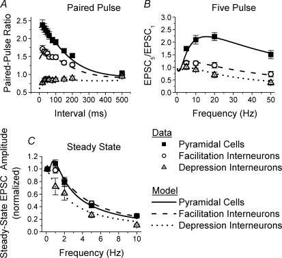
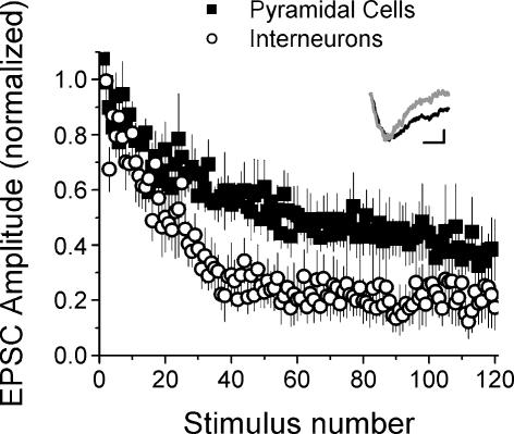
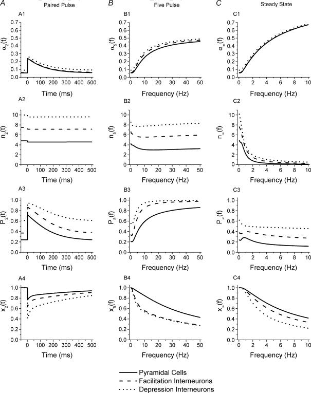
Although it is presynaptic, short-term plasticity has been shown at some synapses to depend upon the postsynaptic cell type. Previous studies have reported conflicting results as to whether Schaffer collateral axons have target-cell specific short-term plasticity. Here we investigate in detail the short-term dynamics of Schaffer collateral excitatory synapses onto CA1 stratum radiatum interneurones versus pyramidal cells in acute hippocampal slices from juvenile rats. In response to three stimulus protocols that invoke different forms of short-term plasticity, we find differences in some but not all forms of presynaptic short-term plasticity, and heterogeneity in the short term plasticity of synapses onto interneurones. Excitatory synapses onto the majority of interneurones had less paired-pulse facilitation than synapses onto pyramidal cells across a range of interpulse intervals (20-200 ms). Unlike synapses onto pyramidal cells, synapses onto most interneurones had very little facilitation in response to short high-frequency trains of five pulses at 5, 10 and 20 Hz, and depressed during trains at 50 Hz. However, the amount of high-frequency depression was not different between synapses onto pyramidal cells versus the majority of interneurones at steady state during 2-10 Hz trains. In addition, a small subset of interneurones (approximately 15%) had paired-pulse depression rather than paired-pulse facilitation, showed only depression in response to the high-frequency five pulse trains, and had more steady-state high-frequency depression than synapses onto pyramidal cells or the majority of interneurones. To investigate possible mechanisms for these differences in short-term plasticity, we developed a mechanistic mathematical model of neurotransmitter release that explicitly explores the contributions to different forms of short-term plasticity of the readily releasable vesicle pool size, release probability per vesicle, calcium-dependent facilitation, synapse inactivation following release, and calcium-dependent recovery from inactivation. Our model fits the responses of each of the three cell groups to the three different stimulus protocols with only two parameters that differ with cell group. The model predicts that the differences in short-term plasticity between synapses onto CA1 pyramidal cells and stratum radiatum interneurones are due to a higher initial release probability per vesicle and larger readily releasable vesicle pool size at synapses onto interneurones, resulting in a higher initial release probability. By measuring the rate of block of NMDA receptors by the open channel blocker MK-801, we confirmed that the initial release probability is greater at synapses onto interneurones versus pyramidal cells. This provides a mechanism by which both the initial strength and the short-term dynamics of Schaffer collateral excitatory synapses are regulated by their postsynaptic target cell.


Figures

Figure 1
Figure 1. Excitatory synapses onto CA1 s. radiatum inhibitory interneurones have less paired-pulse facilitation than synapses onto CA1 pyramidal cells
Examples of EPSCS recorded in response to paired-pulse stimulation of Schaffer collateral axons in a pyramidal cell (A), interneurone with facilitation (B) and interneurone with depression (C). Each trace is the average of 10 responses; traces are overlaid for paired-pulse intervals of 30, 50, 80, 100, 150 and 200 ms. D, group results for paired-pulse ratios (mean ± s.e.m.) from pyramidal cells (squares, n = 32), interneurones with facilitation (circles, n = 57) and interneurones with depression (triangles, n = 9). There are significant differences between three groups (one-way ANOVA, P < 0.05).
Figure 2
Figure 2. Facilitation during short trains is also reduced at excitatory synapses onto interneurones versus pyramidal cells
Examples of EPSCs recorded in response to five-pulse trains in a pyramidal cell (A), interneurone with facilitation (B) and interneurone with depression (C). Each trace is the average of 10 responses; traces are shown for stimulus trains of 5, 20 and 50 Hz. D, group results for five-pulse ratios (EPSC5/EPSC1, mean ± s.e.m.) for pyramidal cells (squares, n = 16), facilitation interneurones (circles, n = 35) and depression interneurones (triangles, n = 8). There are significant differences between the three groups (one-way ANOVA, P < 0.05).
Figure 3
Figure 3. Steady-state high-frequency depression for excitatory synapses onto CA1 pyramidal cells versus those onto CA1 s. radiatum interneurones
Examples of EPSCs recorded at steady state during high-frequency stimulation of Schaffer collateral axon at different frequencies in a pyramidal cell (A), facilitation interneurone (B) and depression interneurone (C). Each trace is the average of 10 responses; traces are shown for steady-state responses to stimulation at 0.1, 5 and 10 Hz. D, example of EPSC amplitudes versus time for stimulation at 2, 5 and 10 Hz to show steady state. Data are from a pyramidal cell; curves have been smoothed by 5 point adjacent averaging. E, group results for steady-state high-frequency depression (mean ± s.e.m., normalized to response size at 0.1 Hz) for pyramidal cells (squares, n = 28), facilitation interneurones (circles, n = 42), and depression interneurones (triangles, n = 6). There is no significant difference between pyramidal cells and facilitation interneurones except at 1 Hz (P > 0.5), but there are significant differences between depression interneurones and the other two cell types (one-way ANOVA, P < 0.05).
Figure 4
Figure 4. Morphology of interneurones does not correlate with the short-term plasticity of their inputs
Paired-pulse ratios obtained from two CA1 s. radiatum interneurones showing different morphological characteristics. A, two biocytin-filled s. radiatum interneurones (interneurones 1 and 2) recorded in the same slice show different morphological characteristics. S. pyramidale is out of the field of view, off the top of the picture. Scale bar, 100 μm. B, paired-pulse ratios are not different at synapses onto interneurone 1 (▪) and interneurone 2 (◯) (P > 0.6).
Figure 5
Figure 5. Model predicts that differences in initial release probability cause the observed changes in short-term plasticity
Mathematical simulations (lines) of experimental data (symbols) provide excellent fits to paired-pulse ratios (A), five-pulse ratios (B), and steady-state high-frequency depression (C) obtained from CA1 pyramidal cells (squares), s. radiatum interneurones with facilitation (circles), and with depression (triangles). For each panel continuous curves are from model fits to pyramidal cell data, dashed lines are from model fits to facilitation interneurone data, and dotted lines are from model fits to depression interneurone data. All curves were calculated using equations from Methods with parameters values given in Table 1 and Table 2. Only the values of the initial vesicular release probability α1 and the initial readily releasable pool size nT are different between the three cell groups; for each cell group, values of α1 and nT are the same for all three stimulus protocols.
Figure 6
Figure 6. Synapses onto interneurones have higher initial release probability as shown by a faster MK-801 blocking rate
Decrease in the NMDA-receptor-mediated EPSC amplitude versus stimulus number in 40 μm MK-801. Rate of block by MK-801 is faster for synapses onto interneurones (◯, mean ± s.e.m., n = 5) versus pyramidal cells (▪, mean ± s.e.m., n = 5, P < 0.005). Inset: examples of average EPSCs in the absence (black line, average of 10 EPSCs from baseline) and presence (grey line, average of first 10 EPSCs in MK-801) of MK-801 for an interneurone. EPSCs have been scaled so their initial peaks match to show the faster decay in the presence of MK-801. Scale bars: 20 ms, 10 pA for control, 5.3 pA in MK-801.
Figure 7
Figure 7. Dynamics of model parameters that govern short-term plasticity for different stimulus patterns
Dynamics of α, the release probability per vesicle (A1–C1); n, the readily releasable vesicle pool size (A2–C2); P, the release probability per active (release-ready) synapse (A3–C3); and x, the fraction of synapses in release-ready state (A4–C4) for different cell types. For each panel continuous curves are from model fits to pyramidal cell data, dashed lines are from model fits to facilitation interneurone data, and dotted lines are from model fits to depression interneurone data. A shows parameter values for the second pulse of paired-pulse stimulation given at different time intervals after the first pulse, which occurs just prior to t= 0. The initial values of the parameters (which govern release on pulse 1) are shown at t < 0. B shows parameter values for the fifth pulse as a function of the frequency of the five-pulse train. C shows parameter values at steady state during constant frequency stimulation at different frequencies. All curves were calculated using equations from Methods with parameter values given in Table 1 and Table 2.

Similar articles

Cited by

References

    1. Allen C, Stevens CF. An evaluation of causes for unreliability of synaptic transmission. Proc Natl Acad Sci U S A. 1994;91:10380–10383. - PMC - PubMed
    1. Augustin I, Rosenmund C, Südhof TC, Brose N. Munc13-1 is essential for fusion competence of glutamatergic synaptic vesicles. Nature. 1999;400:457–461. - PubMed
    1. Bahring R, Bowie D, Benveniste M, Mayer ML. Permeation and block of rat GluR6 glutamate receptor channels by internal and external polyamines. J Physiol. 1997;502:575–589. - PMC - PubMed
    1. Bellingham MC, Lim R, Walmsley B. A novel presynaptic inhibitory mechanism underlies paired pulse depression at a fast central synapse. Neuron. 1999;23:159–170. - PubMed
    1. Bennett MR, Farnell L, Gibson WG. The probability of quantal secretion near a single calcium channel of an active zone. Biophys J. 2000;78:2201–2221. - PMC - PubMed

Publication types

LinkOut - more resources