Comparative Study
. 2005 Aug 30;102(35):12477-82.
doi: 10.1073/pnas.0500584102. Epub 2005 Aug 18.

Dissecting the genetic complexity of human 6p deletion syndromes by using a region-specific, phenotype-driven mouse screen

Affiliations
Comparative Study

Dissecting the genetic complexity of human 6p deletion syndromes by using a region-specific, phenotype-driven mouse screen

Debora Bogani et al. Proc Natl Acad Sci U S A. .

Abstract

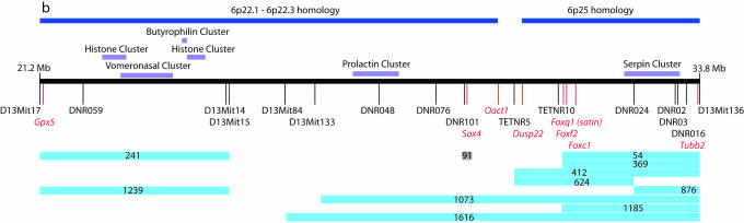
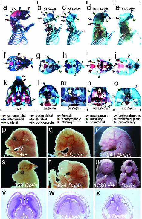
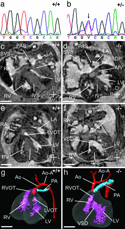
Monosomy of the human chromosome 6p terminal region results in a variety of congenital malformations that include brain, craniofacial, and organogenesis abnormalities. To examine the genetic basis of these phenotypes, we have carried out an unbiased functional analysis of the syntenic region of the mouse genome (proximal Mmu13). A genetic screen for recessive mutations in this region recovered thirteen lines with phenotypes relevant to a variety of clinical conditions. These include two loci that cause holoprosencephaly, two that underlie anophthalmia, one of which also contributes to other craniofacial abnormalities such as microcephaly, agnathia, and palatogenesis defects, and one locus responsible for developmental heart and kidney defects. Analysis of heterozygous carriers of these mutations shows that a high proportion of these loci manifest with behavioral activity and sensorimotor deficits in the heterozygous state. This finding argues for the systematic, reciprocal phenotypic assessment of dominant and recessive mouse mutants. In addition to providing a resource of single gene mutants that model 6p-associated disorders, the work reveals unsuspected genetic complexity at this region. In particular, many of the phenotypes associated with 6p deletions can be elicited by mutation in one of a number of genes. This finding implies that phenotypes associated with contiguous gene deletion syndromes can result not only from dosage sensitivity of one gene in the region but also from the combined effect of monosomy for multiple genes that function within the same biological process.


Figures

Fig. 1.
Fig. 1.
Mutation location. A line diagram of the Del(13)Svea36H region showing human synteny, the position of gene clusters and markers used in the recombination mapping (black) relative to relevant genes in the region (red). The critical interval for each mapped line is shown by the blue line beneath the map. Line 91 carries a mutation in the Sox4 gene.
Fig. 2.
Fig. 2.
Craniofacial and forebrain development. The genotype of mutant animals is represented as Del/m to indicate the mutation (m) in trans to the Del(13)Svea36H chromosome (Del). (a-o) Differential bone and cartilage staining of wild type (+/+; a, f, and k) and mutants from three lines: 54 Del/m (b, c, g, h, l, and m), 1073 Del/m (d, i, and n), and 412 Del/m (e, j, and o). Norma lateralis views (a-e) demonstrate that each mutant specimen is microcephalic and has significant neurocranial, splanchnocranial, and dermatocranial defects, whereas norma basalis views (f-o) highlight the nature of the midline defects of the mutants. The nasal and optic capsules are severely deficient, as are the trabecular basal plate of the neurocranium to which they normally attach and the dermatocranial elements that develop in association with the capsules. Labeled colored arrows represent keys to the identification of homologous elements. (p) Lateral view of the cranial region of a wild-type 15.5-dpc embryo. (q) Lateral view of the cranial region of a line 54 15.5-dpc Del/m embryo showing impaired craniofacial development with anophthalmia, agnathia, and absence of ear pinnae. (r) Lateral view of the cranial region of a line 241 15.5-dpc Del/m embryo showing anophthalmia and absence of ear pinnae. (s) Lateral view of the cranial region of a line 369 15.5-dpc Del/m embryo exhibiting exencephaly. (t) Lateral view of the cranial region of a line 624 15.5 dpc Del/m embryo exhibiting a single, ventrally displaced eye beneath a thin walled holosphere. (u) Anterior view of an 18.5-dpc wild-type embryo (Right) and a 1239 Del/m embryo with cyclopia (Left). (v-x) Transverse sections through the prosencephalon of a 15.5-dpc wild-type embryo (v) or hemizygous mutants (w and x) showing hypoplastic lateral ventricles in lines 412 and 54 with no development of the interhemispheric fissure (arrow in v).
Fig. 3.
Fig. 3.
The Sox4 missense mutation. (a and b) Sequencing of Sox4 in line 91 heterozygotes reveals a T to C transition (arrow) at nucleotide 869. (c-h) Magnetic resonance imaging of the heart in wild-type and Line91 hemizygote mutant embryos at 14.5 dpc. (c) Transverse section through a wild-type embryo at the level of the mitral valve (MV). The left (LV) and right (RV) ventricles are separated by the interventricular septum (IVS) and the left (LA) and right (RA) atria by the primary atrial septum (PAS). The systemic venous sinus (SVS) draining into the RA is indicated. (d) A corresponding section through a Sox4-/- heart. A primum atrial septal defect (ASDP) is seen at the ventral margin of the PAS, with a common atrioventricular junction. The mitral valve is dysplastic. (e and g) Transverse section at the level of the left ventricular outflow tract (LVOT) and 3D reconstruction of a wild-type embryonic heart at 14.5 dpc. The aorta (Ao) originates from the left ventricle (LV), and the main pulmonary artery (PA) from the right ventricle (RV). The right ventricular outflow tract (RVOT) is indicated. The aortic arch (Ao-A) is the same size as the PA. (f and h) Corresponding transverse section and 3D reconstruction of a Sox4-/- heart at 14.5 dpc. A large ventricular septal defect (VSD) connects the RV and LV. The aorta and pulmonary artery both originate from the right ventricle (double-outlet right ventricle). The Ao-A is smaller than the PA. Axes: D, dorsal; V, ventral; L, left; R, right. (Scale bar, 500 μM.)

References

    1. Palmer, C. G., Bader, P., Slovak, M. L., Comings, D. E. & Pettenati, M. J. (1991) Am. J. Med. Genet. 39, 155-160. - PubMed
    1. Davies, A. F., Mirza, G., Sekhon, G., Turnpenny, P., Leroy, F., Speleman, F., Law, C., van Regemorter, N., Vamos, E., Flinter, F., et al. (1999) Hum. Genet. 104, 64-72. - PubMed
    1. Gould, D. B., Jaafar, M. S., Addison, M. K., Munier, F., Ritch, R., MacDonald, I. M. & Walter, M. A. (2004) BMC Med. Genet. 5, 17. - PMC - PubMed
    1. Mirza, G., Williams, R. R., Mohammed, S., Clark, R., Newbury-Ecob, R., Baldinger, S., Flinter, F. & Ragoussis, J. (2004) Eur. J. Hum. Genet. 12, 718-728. - PubMed
    1. Hong, H. K., Lass, J. H. & Chakravarti, A. (1999) Hum. Mol. Genet. 8, 625-637. - PubMed

Publication types

MeSH terms