. 2005 Aug 30;102(35):12507-12.
doi: 10.1073/pnas.0500397102. Epub 2005 Aug 18.

Phosphorylated FADD induces NF-kappaB, perturbs cell cycle, and is associated with poor outcome in lung adenocarcinomas

Affiliations

Phosphorylated FADD induces NF-kappaB, perturbs cell cycle, and is associated with poor outcome in lung adenocarcinomas

Guoan Chen et al. Proc Natl Acad Sci U S A. .

Abstract

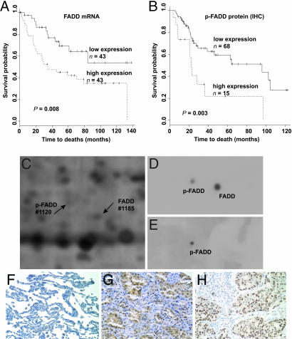
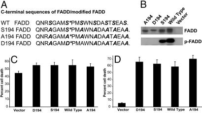
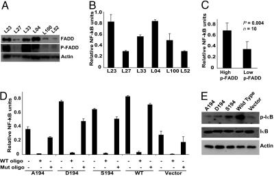
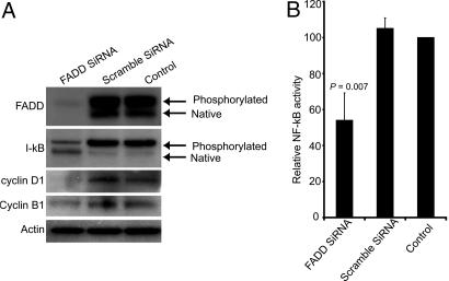
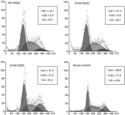
In an effort to identify a clinical biomarker for lung cancer, we used cDNA microarray and 2D protein analyses to demonstrate that increased Fas-associated death domain (FADD) mRNA and protein were significantly associated with poor survival. Analyses of copy number and sequence of the FADD gene in 24 independent tumors ruled out the existence of an amplified and/or mutated FADD gene in aggressive lung cancers. Immunohistochemistry-based tissue microarray analysis showed that nuclear localization of FADD and elevation of the phosphorylated form of FADD (p-FADD) correlated with poor outcome (P = 0.003). Tumors with increased p-FADD expression showed elevated NF-kappaB (P = 0.004) activation, a frequent molecular alteration associated with tumorigenesis and metastasis in a variety of cancers. To provide a link between p-FADD and NF-kappaB, cell culture studies demonstrated that overexpression of p-FADD leads to an increase in NF-kappaB activity and a decrease in the number of cells in the G2 phase of the cell cycle, compared with cells expressing the nonphosphorylatable form of FADD or the vector control. Furthermore, cDNA microarray analyses of lung tumor samples showed that increased levels of FADD transcripts were significantly correlated with overexpression of cyclins D1 (P < 0.01) and B1 (P < 0.01), genes that are involved in the regulation of cell cycle progression and are inducible by NF-kappaB. These studies demonstrate that induction of NF-kappaB activity and its effects on cell-cycle progression may represent a molecular basis underlying the aggressive tumor behavior associated with elevated p-FADD expression in lung adenocarcinoma.


Figures

Fig. 1.
Fig. 1.
FADD mRNA and protein expression in lung adenocarcinomas. (A) Kaplan-Meier survival analysis showed a significantly different outcome between tumors with high and those with low levels of FADD mRNA expression (median value as cutoff value, long-rank test, P = 0.008). (B) Kaplan-Meier survival plot reveals that the 15 patients with highest p-FADD protein expression (score 3 vs. 0 -2) show a poor outcome (P = 0.003) in an independent set of lung adenocarcinomas. (C) Region of a 2D PAGE from a primary lung cancer, showing FADD (1185) and p-FADD (1120) proteins. (D) Western blot with anti-FADD antibody identified two spots representing FADD and p-FADD proteins. (E) Western blot with anti-p-FADD antibody identified only the phosphorylated form of FADD. (F) Immunohistochemical analysis of normal lung tissue, using the anti-FADD antibody on tissue arrays, displays a weak FADD staining. (G) Lung adenocarcinomas display strong cytoplasmic and nuclear immunoreactivity with the anti-FADD antibody. (H) By using the antibody to the p-FADD, immunostaining was predominantly detected in the nucleus of lung adenocarcinomas.
Fig. 2.
Fig. 2.
Generation and characterization of C-terminal FADD mutants. (A) The sequence of the C-terminal domain of FADD harbors key phosphorylation sites at Ser194, as shown by the asterisk. WT FADD contains seven serine residues (italicized) at the C-terminal domain, of which all except Ser194 were replaced by alanine in the mutant FADD constructs. S194, A194, and D194 FADD constructs have serine, alanine, and aspartic acid at position 194. (B) Whole lysates from Jurkat cells expressing the A194 FADD, D194 FADD, S194 FADD, WT FADD, or vector control were examined by Western blotting using either anti-FADD or anti-p-FADD antibodies. (C) Apoptosis induced by 5-fluorouracil in Jurkat cells expressing WT or FADD mutants, monitored by the live-dead assay using calcein AM and PI staining. (D) Cell death, in stable Jurkat cell lines of WT or FADD mutants, induced by activation of the Fas receptor with anti-Fas antibodies was assessed by using calcein AM and PI staining. All of the above-mentioned experiments were performed in at least triplicate and the results plotted as mean (±SEM) of percent cell death.
Fig. 3.
Fig. 3.
Induction of NF-κB activity by p-FADD. (A) Lysates from lung tumor samples expressing high (L23, L33, and L04) and low (L27, L100, and L52) p-FADD were subjected to Western blot analysis using anti-FADD, anti-p-FADD, and anti-actin antibodies. (B) Lysates from nuclei-enriched fractions were prepared from high (L23, L33, and L04) and low (L27, L100, and L52) p-FADD-expressing tumors samples, and NF-κB activity was monitored by using the functional NF-κB TransAM kit from Active Motif. Data shown is derived from mean (±SEM) of relative NF-κB of three separate experiments performed in triplicate. (C) NF-κB activity was significantly higher in tissues containing high levels of p-FADD, as compared with those having low levels of p-FADD (data derived from A and B; n = 10, P = 0.004). (D) Nuclear lysates from various Jurkat cell lines expressing FADD or mutant FADDs were prepared, and NF-κB activity was monitored. For competition experiments, lysates were preincubated with either WT NF-κB consensus oligonucleotide or mutant NF-κB oligonucleotide, and NF-κB activity was then assessed. (E) Western blot analysis of whole-cell lysates obtained from the WT FADD or various mutant FADD-expressing Jurkat cell lines by using anti-I-κB, anti-phospho I-κB, and anti-actin antibodies.
Fig. 4.
Fig. 4.
siRNA mediated down-regulation of FADD results in decrease in NF-κB activity. (A) SKLU-1 lysates from cells transfected with FADD siRNA and scramble siRNA and nontransfected control cells were subjected to Western blot analyses using antibodies to FADD, I-κB, and cyclins D1 and B1. Antibody to actin was used as a control. (B) Nuclear lysates were made from SKLU-1 cells transfected with FADD siRNA and scramble siRNA and from nontransfected control cells and were subjected to NF-κB activity by using the TransAM NF-κB kit from Active Motif. Data shown is derived from mean (±SD) of relative NF-κB activity to nontransfected controls of three separate experiments, performed in triplicate
Fig. 5.
Fig. 5.
Influence of FADD phosphorylation on cell-cycle progression. Analysis of cell-cycle progression of stable Jurkat cell lines expressing the WT FADD or FADD mutants was performed by using FACS. More cells were arrested in G2/M phase in A194 FADD (27.2%) or vector (17.2%), as compared with D194 FADD (5.9%) or WT FADD (3.3%).

References

    1. Beer, D. G., Kardia, S. L., Huang, C. C., Giordano, T. J., Levin, A. M., Misek, D. E., Lin, L., Chen, G., Gharib, T. G., Thomas, D. G., et al. (2002) Nat. Med. 8, 816-824. - PubMed
    1. Chen, G., Gharib, T. G., Wang, H., Huang, C. C., Kuick, R., Thomas, D. G., Shedden, K. A., Misek, D. E., Taylor, J. M., Giordano, T. J., et al. (2003) Proc. Natl. Acad. Sci. USA 100, 13537-13542. - PMC - PubMed
    1. Chinnaiyan, A. M., O'Rourke, K., Tewari, M. & Dixit, V. M. (1995) Cell 81, 505-512. - PubMed
    1. Boldin, M. P., Varfolomeev, E. E., Pancer, Z., Mett, I. L., Camonis, J. H. & Wallach, D. (1995) J. Biol. Chem. 270, 7795-7798. - PubMed
    1. Zhang, J. & Winoto, A. (1996) Mol. Cell. Biol. 16, 2756-2763. - PMC - PubMed

Publication types

MeSH terms