

Brain morphometry using diffusion-weighted magnetic resonance imaging: application to schizophrenia
- PMID: 16110271
- PMCID: PMC1539168
- DOI: 10.1097/01.wnr.0000177001.27569.06
Brain morphometry using diffusion-weighted magnetic resonance imaging: application to schizophrenia
Abstract
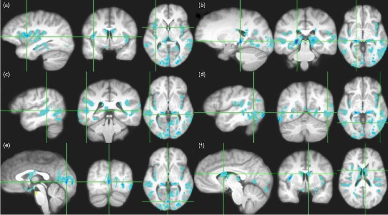
Loss of cortical gray matter is accompanied by a commensurate increase in the sulcal and intraventricular cerebrospinal fluid volume. On diffusion-weighted magnetic resonance imaging, this would be reflected as a higher apparent diffusion coefficient in affected brain regions. On the basis of the above premise, we suggest that the apparent diffusion coefficient may be used as a surrogate marker for the assessment of regional brain volume deficits. We demonstrate this approach by voxelwise analysis of registered apparent diffusion coefficient images from a group of 15 patients with schizophrenia and 15 age-matched healthy controls. We found widespread regional apparent diffusion coefficient increases in patients. Affected areas included the bilateral insular cortex, hippocampus, temporal lobe, and occipital areas. These results largely concur with previous findings of cortical volume deficits in schizophrenia.
Figures
References
-
- Palmen SJ, van Engeland H. Review on structural neuroimaging findings in autism. J Neural Transm. 2004;111:903–929. - PubMed
-
- Bogerts B, Lieberman JA, Ashtari M, Bilder RM, Degreef G, Lerner G, et al. Hippocampus–amygdala volumes and psychopathology in chronic schizophrenia. Biol Psychiatry. 1993;33:236–246. - PubMed
-
- Takahashi T, Suzuki M, Hagino H, Zhou SY, Kawasaki Y, Nohara S, et al. Bilateral volume reduction of the insular cortex in patients with schizophrenia: a volumetric MRI study. Psychiatry Res. 2004;132:187–196. - PubMed
-
- Davis KA, Kwon A, Cardenas VA, Deicken RF. Decreased cortical gray and cerebral white matter in male patients with familial bipolar I disorder. J Affect Disord. 2004;82:475–485. - PubMed
Publication types
MeSH terms
Grants and funding
LinkOut - more resources
Full Text Sources
Medical
