. 2005 Sep;115(9):2412-22.
doi: 10.1172/JCI24086. Epub 2005 Aug 18.

ATP and purinergic receptor-dependent membrane traffic in bladder umbrella cells

Affiliations

ATP and purinergic receptor-dependent membrane traffic in bladder umbrella cells

Edward C Y Wang et al. J Clin Invest. 2005 Sep.

Abstract

The umbrella cells that line the bladder are mechanosensitive, and bladder filling increases the apical surface area of these cells; however, the upstream signals that regulate this process are unknown. Increased pressure stimulated ATP release from the isolated uroepithelium of rabbit bladders, which was blocked by inhibitors of vesicular transport, connexin hemichannels, ABC protein family members, and nucleoside transporters. Pressure-induced increases in membrane capacitance (a measure of apical plasma membrane surface area where 1 microF approximately equals 1 cm2) were inhibited by the serosal, but not mucosal, addition of apyrase or the purinergic receptor antagonist PPADS. Upon addition of purinergic receptor agonists, increased capacitance was observed even in the absence of pressure. Moreover, knockout mice lacking expression of P2X2 and/or P2X3 receptors failed to show increases in apical surface area when exposed to hydrostatic pressure. Treatments that prevented release of Ca2+ from intracellular stores or activation of PKA blocked ATPgammaS-stimulated changes in capacitance. These results indicate that increased hydrostatic pressure stimulates release of ATP from the uroepithelium and that upon binding to P2X and possibly P2Y receptors on the umbrella cell, downstream Ca2+ and PKA second messenger cascades may act to stimulate membrane insertion at the apical pole of these cells.


Figures

Figure 1
Figure 1
ATP localization, release, and hydrolysis in uroepithelium. (A and B) Pressure-induced release of ATP from the serosal (A) or mucosal (B) surfaces of rabbit uroepithelium. The epithelial tissue was mounted in modified Ussing stretch chambers, equilibrated, and pretreated with the indicated drug 15 minutes before the start of the experiment. ATP release was measured just prior to the start of the experiment (background [BKG]), immediately following the rise in hydrostatic pressure (t = 0), and 3, 6, 10, 20, 30, 50, and 60 minutes after the pressure was raised. Results are expressed as mean ± SEM (n ≥ 3). *Statistically significant difference (P < 0.05) relative to the appropriate control. (C) Rabbit uroepithelium was incubated with 5 μM quinacrine for 30 minutes at room temperature, subsequently labeled with FM4-64 (to label cell membranes), and then imaged using a confocal microscope. The image is a projection of the Z series. Quinacrine staining is shown in green, and FM4-64 staining is shown in blue. (D) ATP (50 μM) was added to the mucosal or serosal hemichamber (labeled “Mucosal” and “Serosal,” respectively) of tissue mounted in Ussing stretch chambers. In control reactions, tissue was replaced with plastic film (labeled “Mucosal control” and “Serosal control”). At the designated time points, samples were taken, and the ATP concentration remaining in the hemichamber was measured. Results are expressed as mean ± SEM (n ≥ 3). *Statistically significant difference (P < 0.05) relative to the appropriate control.
Figure 2
Figure 2
Effects of ATP, apyrase, and PPADS on changes in membrane capacitance. (A and B) Isolated rabbit uroepithelial tissue was exposed to pressure alone (Pressure), pressure and 0.4 U/ml apyrase (Apyrase + pressure), or pressure and 100 μM PPADS (PPADS + pressure). PPADS and apyrase were added to either the serosal (A) or mucosal (B) surfaces of the tissue. The mean changes in capacitance ± SEM (n ≥ 3) are shown. *Statistically significant difference (P < 0.05) relative to the samples treated with pressure alone. (C) ATP (at the indicated concentration) was added to the serosal surface of isolated rabbit uroepithelium without pressure stimulus. The mean change (± SEM) in capacitance at 300 minutes after addition of ATP was recorded (n ≥ 3) and plotted versus the log of the ATP concentration.
Figure 3
Figure 3
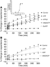
Modulation of membrane capacitance by serosal addition of purinergic receptor agonists. (A and B) ATP or the following ATP agonists were added at a final concentration of 50 μM to the serosal surface of isolated rabbit uroepithelium without exposure to pressure changes: ATPγS, 2MeSATP, benzoyl ATP (BzATP), UTP, ADP, or 2MeSADP. The inset in A shows a dose-response curve for the change in capacitance recorded 300 minutes after addition of the specified concentration of ATPγS. Data are shown as mean changes in capacitance ± SEM (n ≥ 3). *Statistically significant difference (P < 0.05) relative to untreated control preparations.
Figure 4
Figure 4
ATPγS-stimulated apical endocytosis in bladder umbrella cells. The apical surface of rabbit umbrella cells was biotinylated and then incubated for 0, 5, 15, 30, 60, or 120 minutes in the absence (Control) or presence of 50 μM ATPγS added to the serosal surface of the uroepithelium. In samples treated with ATPγS, the t = 0 sample was taken immediately after addition of the agonist. The fraction of MESNA-protected biotinylated proteins was detected by probing Western blots with streptavidin-HRP, and the blots were quantified by densitometry. Data are expressed as mean ± SEM (n ≥ 5). *Statistically significant difference (P < 0.05) relative to the appropriate control. The control data was published previously (26).
Figure 5
Figure 5
Localization of P2X3 in the uroepithelium and responses to pressure changes in P2X2- and P2X3-knockout mice. (A and B) Localization of P2X3 in cryosections of rabbit bladder uroepithelium. (A) P2X3 staining is shown in green, and the umbrella cells (UC) are marked with arrows. (B) Composite image with P2X3 staining shown in green, rhodamine-phalloidin–labeled actin in red, and Topro-3–labeled (Molecular Probes) nuclei in blue. (C) Whole mounted rabbit uroepithelium showing the distribution of the nuclei (blue) and P2X3 (green). The image is a 3-dimensional reconstruction of a Z series collected with a confocal microscope. The image was tilted around the x axis to emphasize the 3-dimensional aspect of the image. The grid is a 3-dimensional scale bar, with each side of the square approximately equivalent to 12.5 μm. (D) Bladders from mice of the indicated strains were mounted in Ussing stretch chambers, the pressure was increased at t = 0, and the capacitance was recorded. Data shown are mean ± SEM (n ≥ 3). *Statistically significant difference (P < 0.05) relative to the appropriate control.
Figure 6
Figure 6
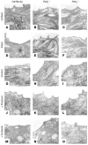
Transmission electron microscopy (TEM) analysis of apical pole of umbrella cells in wild-type and knockout mice. (AF) C57BL/6J wild-type (A and D), P2X2_/_ (B and E), or P2X3_/_ (C and F) mice were catheterized, and the urine content of the bladder was drained. After a 30-minute incubation, the bladders were either excised and processed for TEM (AC), or the bladder was slowly filled with normal saline at a rate of 1.4 μl/min for 180 minutes (DF). Examples of fusiform vesicles (FV) are marked in A. (GO) TEM analysis of the apical poles of umbrella cells from C57BL/6J wild-type (G, J, and M), P2X2_/_ (H, K, and N), or P2X3_/_ (I, L, and O) mice mounted in Ussing stretch chambers and incubated in the absence of hydrostatic pressure (GI) or the presence of hydrostatic pressure (JL) or treated with 10 μM forskolin (added to the mucosal hemichamber) in the absence of pressure (MO).
Figure 7
Figure 7
Potential role of Ca2+ and PKA in ATPγS-stimulated changes in membrane capacitance. Rabbit uroepithelium was mounted in modified Ussing chambers and preincubated with 75 μM 2-aminoethoxydiphenylborate (APB), 50 μM ryanodine, or 10 μM H89 for 30 minutes as indicated. In the Ca2+-free experiments, the normal Krebs solution was isovolumetrically replaced with Krebs solution lacking Ca2+. At t = 0, 50 μM ATPγS was added into the serosal hemichamber, and changes in capacitance were monitored over time in the continued presence of the appropriate drug. Data are mean ± SEM (n ≥ 5). *Statistically significant difference (P < 0.05) relative to the ATPγS reaction.
Figure 8
Figure 8
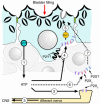
Model for ATP and purinergic receptor–regulated exocytosis/endocytosis in bladder umbrella cells. The accumulation of urine in the bladder increases hydrostatic pressure, stimulating release of ATP from uroepithelium (step 1) and possibly other adjacent cell types (not shown). ATP release may occur through multiple mechanisms, including vesicular release, conductance of ATP though ABC or nucleoside transporters, or movement of ATP across connexin hemichannels. The serosally released ATP may bind to P2X3-containing receptors present on afferent nerve processes (step 2), increasing nerve firing and relaying bladder filling to the CNS (step 3). Serosally released ATP may also bind to receptors containing P2X2 and/or P2X3 subunits present on the uroepithelium, including the basolateral surface of the umbrella cell layer (step 4). Other receptors, including P2X or P2Y receptor isoforms, are also present on the serosal surface of the epithelium and may also be stimulated by ATP (or other released or processed nucleotides/nucleosides). Ligand binding causes increased levels of cytoplasmic Ca2+ (a result of Ca2+ influx from outside the cell and efflux from intracellular stores) and activation of PKA that induces apical membrane turnover including discoidal/fusiform vesicle exocytosis and endocytosis (step 5). Although in the simplest model, ATP binds to purinergic receptors on the umbrella cell layer, it remains possible that ATP binds to purinergic receptors present on basal/intermediate cells (step 6), or other cell types, to signal the release of unidentified secretagogues that act upon umbrella cells to stimulate vesicle exocytosis (step 7). For simplicity, mucosal purinergic signaling events are not shown; however, these could be important in modulating the signaling pathways that initiate at the serosal surface of the umbrella cells.

Similar articles

Cited by

References

    1. Novak I. ATP as a signaling molecule: the exocrine focus [review] News Physiol. Sci. 2003;18:12–17. - PubMed
    1. Burnstock G. Purine-mediated signalling in pain and visceral perception. Trends Pharmacol. Sci. 2001;22:182–188. - PubMed
    1. Cooke HJ, Wunderlich J, Christofi FL. “The force be with you”: ATP in gut mechanosensory transduction [review] News Physiol. Sci. 2003;18:43–49. - PubMed
    1. Sorensen CE, Novak I. Visualization of ATP release in pancreatic acini in response to cholinergic stimulus. Use of fluorescent probes and confocal microscopy. J. Biol. Chem. 2001;276:32925–32932. - PubMed
    1. Maroto R, Hamill OP. Brefeldin A block of integrin-dependent mechanosensitive ATP release from Xenopus oocytes reveals a novel mechanism of mechanotransduction. J. Biol. Chem. 2001;276:23867–23872. - PubMed

Publication types

MeSH terms