. 2005 Sep;115(9):2393-401.
doi: 10.1172/JCI25437. Epub 2005 Aug 18.

TRPA1 induced in sensory neurons contributes to cold hyperalgesia after inflammation and nerve injury

Affiliations

TRPA1 induced in sensory neurons contributes to cold hyperalgesia after inflammation and nerve injury

Koichi Obata et al. J Clin Invest. 2005 Sep.

Erratum in

  • J Clin Invest. 2010 Jan;120(1):394

Abstract

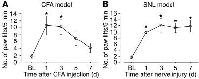
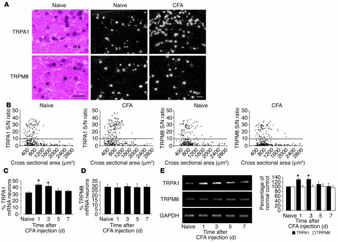
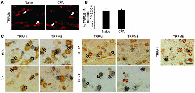
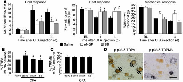
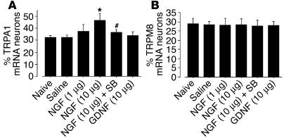
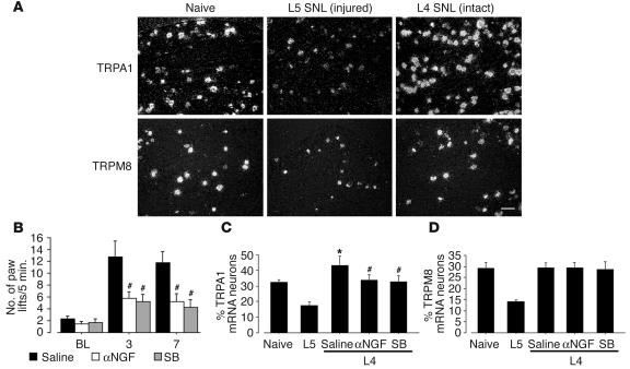
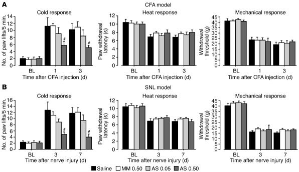
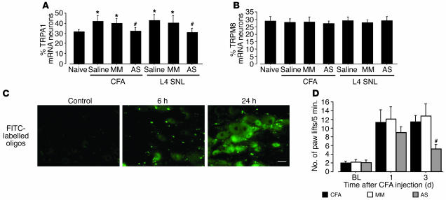
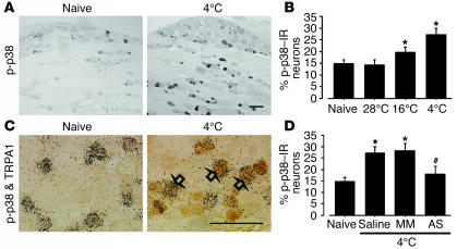
Cold hyperalgesia is a well-documented symptom of inflammatory and neuropathic pain; however, the underlying mechanisms of this enhanced sensitivity to cold are poorly understood. A subset of transient receptor potential (TRP) channels mediates thermosensation and is expressed in sensory tissues, such as nociceptors and skin. Here we report that the pharmacological blockade of TRPA1 in primary sensory neurons reversed cold hyperalgesia caused by inflammation and nerve injury. Inflammation and nerve injury increased TRPA1, but not TRPM8, expression in tyrosine kinase A-expressing dorsal root ganglion (DRG) neurons. Intrathecal administration of anti-nerve growth factor (anti-NGF), p38 MAPK inhibitor, or TRPA1 antisense oligodeoxynucleotide decreased the induction of TRPA1 and suppressed inflammation- and nerve injury-induced cold hyperalgesia. Conversely, intrathecal injection of NGF, but not glial cell line-derived neurotrophic factor, increased TRPA1 in DRG neurons through the p38 MAPK pathway. Together, these results demonstrate that an NGF-induced TRPA1 increase in sensory neurons via p38 activation is necessary for cold hyperalgesia. Thus, blocking TRPA1 in sensory neurons might provide a fruitful strategy for treating cold hyperalgesia caused by inflammation and nerve damage.


Figures

Figure 1
Figure 1
Time course of the exaggerated response to cold after peripheral inflammation and nerve injury. (A and B) The number of paw lifts on a cold plate at 5°C for the 5-minute testing period was examined at days 1, 3, 5, and 7 after CFA injection (A) and L5 SNL (B). Data represent mean ± SEM; n = 8 per group. BL, baseline. *P < 0.05 compared with the naive control.
Figure 2
Figure 2
Marked upregulation of TRPA1, but not TRPM8, mRNA in DRG neurons after peripheral inflammation induced by CFA. (A) Bright- and dark-field photomicrographs of ISHH showing expression of TRPA1 and TRPM8 mRNA in the naive DRG and the ipsilateral DRG at day 1 after inflammation. Scale bars: 100 μm. (B) Scatterplot diagrams made by plotting the individual cell profiles at day 1 after CFA injection. The gray lines represent the borderlines between the negatively and positively labeled neurons (signal/noise [S/N] ratio = 10). (C and D) Time course of the mean percentages of TRPA1 (C) and TRPM8 (D) mRNA–positive neurons after CFA injection. (E) mRNA expression of TRPA1 and TRPM8 in the DRG after inflammation, as detected by RT-PCR. Quantification of RT-PCR data is shown at right. Data represent mean ± SD; n = 4 per group. *P < 0.05 compared with the naive control.
Figure 3
Figure 3
No change of TRPM8 protein in the DRG and no overlap between TRPM8- and TRPA1-expressing neurons after inflammation. (A) Protein expression of TRPM8 in the naive DRG and the ipsilateral DRG at day 3 after inflammation, as detected by immunohistochemistry. TRPM8-immunoreactive (TRPM8-IR) neurons were invariably small or medium in size (arrows). (B) Quantification of the percentage of TRPM8-IR neurons at day 3 after CFA injection. (C) Double labeling by a combined method of ISHH and immunohistochemistry for TRPA1 or TRPM8 mRNA and trkA-IR, SP-IR, CGRP-IR, and TRPV1-IR in the DRG at day 3 after CFA injection. Double labeling for TRPA1 and TRPM8 mRNA by dual ISHH is shown at right. TRPA1- and TRPM8-expressing neurons were clearly distinguishable. Open arrows indicate double-labeled neurons. Scale bars: 50 μm.
Figure 4
Figure 4
Anti-NGF and a p38 MAPK inhibitor reverse cold hyperalgesia and TRPA1 upregulation induced by inflammation. (A) Cold and heat hyperalgesia were tested using the cold plate test and the plantar test, respectively, at days 1 and 3 after CFA injection. Mechanical allodynia was determined with a Dynamic Plantar Aesthesiometer. Data represent mean ± SEM; n = 8 per group. (B and C) Quantification of the percentages of TRPA1 (B) and TRPM8 (C) mRNA–positive neurons at day 3 after CFA injection. Data represent mean ± SD; n = 4 per group. (D) Double labeling for TRPA1 or TRPM8 mRNA and p-p38–IR at day 3 after CFA injection. Open arrows indicate double-labeled neurons. SB, SB203580. *P < 0.05 compared with the naive control; #P < 0.05 compared with the vehicle group. Scale bar: 50 μm.
Figure 5
Figure 5
NGF, but not GDNF, induces an increase of TRPA1 expression in DRG neurons via p38 MAPK activation. Quantification of the percentage of TRPA1 (A) and TRPM8 (B) mRNA–positive neurons at day 3 after the injection. Data represent mean ± SD; n = 4 per group. *P < 0.05 compared with the naive control. #P < 0.05 compared with the NGF 10-μg group.
Figure 6
Figure 6
Anti-NGF and a p38 MAPK inhibitor suppress cold hyperalgesia and TRPA1 upregulation in the spared L4 DRG neurons caused by injury to the L5 spinal nerve. (A) Dark-field photomicrograph of ISHH showing the expression of TRPA1 and TRPM8 mRNA in the naive DRG, the injured L5 DRG, and the intact L4 DRG at day 7 after L5 SNL. (B) Cold hyperalgesia was determined using the cold plate test at days 3 and 7 after L5 SNL. Data represent mean ± SEM; n = 8 per group. (C and D) Quantification of the percentage of TRPA1 (C) and TRPM8 (D) mRNA–positive neurons at day 7 after surgery. Data represent mean ± SD; n = 4 per group. *P < 0.05 compared with the naive control; #P < 0.05 compared with the vehicle group. Scale bar: 100 μm.
Figure 7
Figure 7
Reversal of hyperalgesia to cold by a selective blockade of TRPA1 expression. (A and B) Pretreatment with TRPA1 AS-ODN (0.5 nmol/μl–1/h–1) (AS 0.50) prevents cold hyperalgesia, but not heat hyperalgesia or mechanical allodynia, induced by peripheral inflammation (A) and injury to the L5 spinal nerve (B). Cold and heat hyperalgesia were tested using the cold plate test and the plantar test, respectively, at days 1 and 3 after CFA injection or at days 3 and 7 after L5 SNL. Mechanical allodynia was examined using a Dynamic Plantar Aesthesiometer. Data represent mean ± SEM; n = 8 per group. #P < 0.05 compared with the MM-ODN (0.5 nmol/μl–1/h–1) (MM 0.50) group.
Figure 8
Figure 8
Confirmation of a selective blockade of TRPA1 expression in a time-dependent manner. (A and B) TRPA1 AS-ODN (0.5 nmol/μl–1/h–1) attenuates the induction of TRPA1, but not TRPM8, mRNA induced by peripheral inflammation and injury to the L5 spinal nerve. Quantification of the percentage of TRPA1 (A) and TRPM8 (B) mRNA–positive neurons at day 3 after CFA injection or in the L4 DRG at day 7 after L5 SNL. Data represent mean ± SD; n = 4 per group. (C) Fluorescence in the naive DRG and the DRG at 6 and 24 hours after intrathecal injection of FITC-labeled ODN. Scale bar: 50 μm. (D) Effect of treatment with TRPA1 AS-ODN (0.5 nmol/μl–1/h–1) following CFA injection on CFA-induced cold hyperalgesia at days 1 and 3. Data represent mean ± SEM; n = 8 per group. *P < 0.05 compared with naive control. #P < 0.05 compared with MM-ODN (0.5 nmol/μl–1/h–1) group.
Figure 9
Figure 9
Activation of p38 MAPK in TRPA1-containing neurons by noxious cold stimulation. (A) p-p38 labeling in naive DRG and ipsilateral DRG 2 minutes after cold stimulation at 4°C. (B) Quantification of the percentage of p-p38–IR neurons after innocuous and noxious cold stimulation. (C) Double labeling for TRPA1 mRNA and p-p38–IR in naive DRG and ipsilateral DRG 2 minutes after cold stimulation at 4°C. Open arrows indicate double-labeled neurons. (D) Effect of pretreatment with TRPA1 AS-ODN (0.5 nmol/μl–1/h–1) on the percentage of p-p38–IR neurons after cold stimulation at 4°C. Data represent mean ± SD; n = 4 per group. *P < 0.05 compared with naive control. #P < 0.05 compared with MM-ODN (0.5 nmol/μl–1/h–1) group. Scale bars: 100 μm.

References

    1. LaMotte RH, Thalhammer JG. Response properties of high-threshold cutaneous cold receptors in the primate. Brain Res. 1982;244:279–287. - PubMed
    1. Kress M, Koltzenburg M, Reeh PW, Handwerker HO. Responsiveness and functional attributes of electrically localized terminals of cutaneous C-fibers in vivo and in vitro. J. Neurophysiol. 1992;68:581–595. - PubMed
    1. Hao JX, Yu W, Xu XJ, Wiesenfeld-Hallin Z. Capsaicin-sensitive afferents mediate chronic cold, but not mechanical, allodynia-like behavior in spinally injured rats. Brain Res. 1996;722:177–180. - PubMed
    1. Hama AT. Capsaicin-sensitive primary afferents mediate responses to cold in rats with a peripheral mononeuropathy. Neuroreport. 2002;13:461–464. - PubMed
    1. Jordt SE, McKemy DD, Julius D. Lessons from peppers and peppermint: the molecular logic of thermosensation. Curr. Opin. Neurobiol. 2003;13:487–492. - PubMed

Publication types

MeSH terms