

Review
doi: 10.1002/dvdy.20521.
Challenges in the study of neuronal differentiation: a view from the embryonic eye
Affiliations
- PMID: 16110510
- PMCID: PMC1373677
- DOI: 10.1002/dvdy.20521
Item in Clipboard
Review
Challenges in the study of neuronal differentiation: a view from the embryonic eye
Dev Dyn.
2005 Nov.
Abstract
Progress in the study of the molecular mechanisms that regulate neuronal differentiation has been quite impressive in recent years, and promises to continue to an equally fast pace. This should not lead us into a sense of complacency, however, because there are still significant barriers that cannot be overcome by simply conducting the same type of experiments that we have been performing thus far. This article will describe some of these challenges, while highlighting the conceptual and methodological breakthroughs that will be necessary to overcome them.
Developmental Dynamics 234:454-463, 2005. (c) 2005 Wiley-Liss, Inc.
Figures
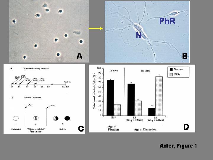
. In vitro analysis of progenitor cell “commitment”. A) Morphologically homogeneous population of progenitor cells isolated from the chick embryo retina before the onset of overt differentiation. The cells are grown at low density, to minimize contact-mediated cell interactions. B) After several days in culture, some of the progenitor cells differentiate as photoreceptors (PhR), while others differentiate as non-photoreceptor, predominantly amacrine neurons (N). C) Diagrammatic summary of the window-labeling technique, which allows identifying cells that undergo terminal mitosis during narrowly defined periods of time. As shown in the top panel, the technique involves the sequential administration of tritiated thymidine (3HT) followed 5 hr later by an initial injection of bromodeoxyuridine (BrDU), which is repeated at daily intervals. As shown in the bottom panel, cells that are already postmitotic at the time of 3HT injection appear unlabeled, and cells that divide once or several times after BrDU administration are BrDU(+); the only cells that are 3HT(+)/BrDU(-) are those that are in S-phase during the 5 hr time interval between 3HT and BrDU administration, and became postmitotic immediately thereafter. D) Analysis of the fate of cells born during a 5 hr interval on embryonic day 5 (WL5). When the cells are allowed to develop in vivo until embryonic day 18, nearly 75% of the cells differentiate as non-photoreceptor neurons, and only 25% differentiate as photoreceptors. Similar results are seen when the cells are isolated for culture on embryonic day 8, after 72 hr of exposure to the retinal microenvironment. On the other hand, when the WL5 cells are exposed to the in vivo microenvironment for only 24 hr before isolation, they give rise predominantly to photoreceptors. As discussed in the text, the experiments indicate that the cells remain plastic after their terminal mitosis. Panels A and B from Madreperla and Adler (1989); panels B and C from Belecky-Adams et al. (1996), reprinted with permission from Elsevier, Inc.
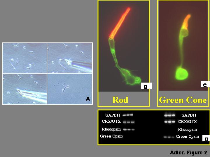
. Analysis of gene expression in isolated retinal cells. A) Retinas are dissociated and individual cells captured as described by Wahlin et al. (2004). B,C) Rod and cone photoreceptor cells isolated from chick embryo retinas can be identified by morphological criteria, verified by immunocytochemistry with antibodies against the visual pigments expressed in each cell type. Individual cells can be processed for cDNA synthesis and exponential amplification using protocols described by Brady and Iscove (1993), and Dulac (1998). D) The identity of the cells can be further investigated by PCR amplification of housekeeping genes (e.g., GAPDH), general photoreceptor markers (e.g., CRX/OTX), and visual pigments which are selectively expressed in individual photoreceptor types (e.g., rhodopsin in rods, green opsin in green cones).
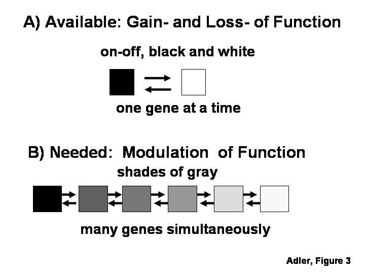
A) There are now many methods for gain- and loss-of function experiments, which allow targeting only one or two genes at a time, and do so in a “black-or-white”, all-or-none manner. B) These methods appear insufficient for the analysis of combinatorial mechanisms regulating cell differentiation, which would require methods for modulating the level of expression of groups of genes.
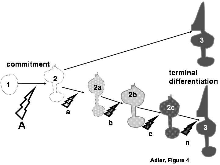
Molecules that are first expressed during the terminal differentiation of a cell type (cell # 3) are frequently used as “markers” to evaluate the inductive signals (A) through which an undifferentiated progenitor cell (cell # 1) becomes committed to a particular differentiated lineage (cell # 2). Such use would only be justified if the entire process of cell differentiation is controlled cell-autonomously by intrinsic mechanisms set in motion at the time of progenitor cell commitment (top pathway). On the other hand, the approach would not be warranted if additional inductive signals (bottom pathway, a, b, c, n) re necessary for a committed progenitor to reach terminal differentiation through a series of intermediate stages (2a, 2b, 2c).
References
-
- Abe Y, Minegishi T, Leung PC. Activin receptor signaling. Growth Factors. 2004;22:105–110. - PubMed
-
- Adler R. A model of retinal cell differentiation in the chick embryo. Prog Ret Eye Res. 2000;20:529–557. - PubMed
-
- Adler R, Belecky-Adams TL. The role of bone morphogenetic proteins in the differentiation of the ventral optic cup. Development. 2002;129:3161–3171. - PubMed
-
- Adler R, Curcio C, Hicks D, Price D, Wong F. Cell death in age-related macular degeneration. Mol Vis. 1999;5:31. - PubMed
Publication types
MeSH terms
Grants and funding
LinkOut - more resources
Full Text Sources
Other Literature Sources
