

Multiple myelomas in cats
- PMID: 16112593
- PMCID: PMC7130061
- DOI: 10.1016/j.jfms.2004.12.005
Multiple myelomas in cats
Abstract
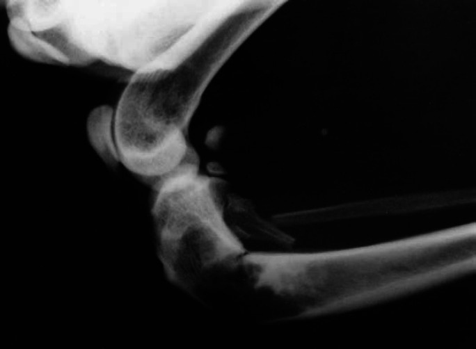
Multiple myelomas are uncommon neoplasms of the bone marrow of cats [Weber NA, Tebeau CS (1998) An unusual presentation of multiple myeloma in two cats. Journal of the American Animal Hospital Association34 (6), 477-483]. Nine cats diagnosed with multiple myelomas were retrospectively identified over a 16-year period (1986-2002). Cats with multiple myelomas were older than 7 years (mean age 11.7 years); six males and three females were affected (2.1), but no breed predisposition was evident. Treatment of multiple myelomas consisted of supportive management in the nine cats and anti-neoplastic therapy in eight cats. Supportive treatment consisted of maintaining hydration, renal function and antimicrobial therapy even when there was no sign of infection. Anti-neoplastic therapy with melphalan and prednisolone was carried out in eight cats. Three failed to respond to treatment and five responded to treatment, but the response was only partial and temporary in one cat. The five cats that responded were improved clinically and had reduced serum protein levels. Five out of eight cats (63%) responded to chemotherapy, and it appeared to be complete in four cats and partial in one cat. Survival time in those cats was 15, 4, 17 and 24 months.
Figures


References
-
- Alexanian R., Dimopouls M. The treatment of multiple myeloma, New England Journal of Medicine 330, 1994, 484–489. - PubMed
-
- Bienzle D., Silverstein D.C., Chaffin K. Multiple myeloma in cats: variable presentation with different immunoglobulin isotypes in two cats, Veterinary Pathology 37 (4), 2000, 364–369. - PubMed
-
- Binta M.G., Obwolo M.J., Ndikuwera J., Mushi E.Z., Hill F.W.G., Rogers S.E. Multiple myeloma associated with paraproteinuria and hyperviscosity syndrome: a case report, Zimbabwe Veterinary Journal 26 (3–4), 1995, 102, 20 ref
-
- Dunn T.B. Plasma cell neoplasms beginning in the ileocecal area in strain C34 mice, Journal of the National Cancer Institute 19, 1957, 371–391. - PubMed
-
- Eastman C.A. Plasma cell tumours in a cat, Feline Practice 24, 1996, 26.
Publication types
MeSH terms
Substances
LinkOut - more resources
Full Text Sources
Medical
Miscellaneous
