

Characterization and application of monoclonal antibodies against N protein of SARS-coronavirus
- PMID: 16112641
- PMCID: PMC7092910
- DOI: 10.1016/j.bbrc.2005.08.032
Characterization and application of monoclonal antibodies against N protein of SARS-coronavirus
Abstract
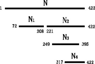
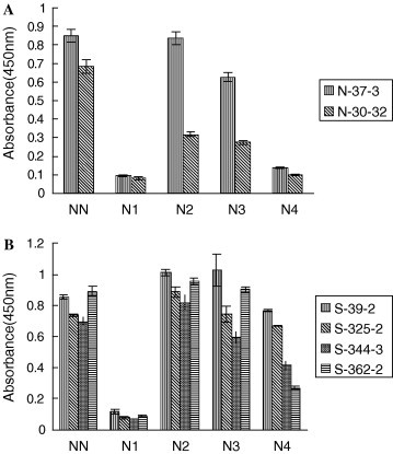
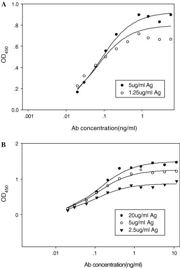
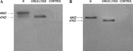
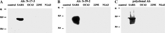
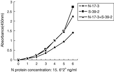
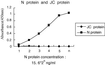
Severe acute respiratory syndrome-coronavirus (SARS-CoV) causes an infectious disease through respiratory route. Diagnosing the disease effectively and accurately at early stage is essential for preventing the disease transmission and performing antiviral treatment. In this study, we raised monoclonal antibodies (mAbs) against the nucleocapsid (N) protein of SARS-CoV and mapped epitopes by using different truncated N protein fragments. The mapping of those epitopes was valuable for constructing pair-Abs used in serological diagnosis. The results showed that all of the six raised mAbs were divided into two groups recognizing the region of amino acids 249-317 (A group) or 317-395 (B group). This region spanning amino acids 249-395 contains predominant B cell epitopes located at the C-terminus of N protein. One pair-Abs, consisting of N protein-specific rabbit polyclonal antibody and SARS-CoV N protein-specific mAb, was selected to construct a sandwich ELISA-kit. The kit was able to specifically detect SARS-CoV N proteins in serum samples.
Figures
References
-
- Rota P.A., Oberste M.S., Monroe S.S., Nix W.A., Campagnoli R., Icenogle J.P., Penaranda S., Bankamp B., Maher K., Chen M.H., Tong S., Tamin A., Lowe L., Frace M., DeRisi J.L., Chen Q., Wang D., Erdman D.D., Peret T.C., Burns C., Ksiazek T.G., Rollin P.E., Sanchez A., Liffick S., Holloway B., Limor J., McCaustland K., Olsen-Rasmussen M., Fouchier R., Gunther S., Osterhaus A.D., Drosten C., Pallansch M.A., Anderson L.J., Bellini W.J. Characterization of a novel coronavirus associated with Severe Acute Respiratory Syndrome. Science. 2003;300(5624):1394–1399. - PubMed
-
- Marra M.A., Jones S.J., Astell C.R., Holt R.A., Brooks-Wilson A., Butterfield Y.S., Khattra J., Asano J.K., Barber S.A., Chan S.Y., Cloutier A., Coughlin S.M., Freeman D., Girn N., Griffith O.L., Leach S.R., Mayo M., McDonald H., Montgomery S.B., Pandoh P.K., Petrescu A.S., Robertson A.G., Schein J.E., Siddiqui A., Smailus D.E., Stott J.M., Yang G.S., Plummer F., Andonov A., Artsob H., Bastien N., Bernard K., Booth T.F., Bowness D., Czub M., Drebot M., Fernando L., Flick R., Garbutt M., Gray M., Grolla A., Jones S., Feldmann H., Meyers A., Kabani A., Li Y., Normand S., Stroher U., Tipples G.A., Tyler S., Vogrig R., Ward D., Watson B., Brunham R.C., Krajden M., Petric M., Skowronski D.M., Upton C., Roper R.L. The Genome sequence of the SARS-associated coronavirus. Science. 2003;300(5624):1399–1404. - PubMed
-
- Wu H.S., Hsieh Y.C., Su I.J., Lin T.H., Chiu S.C., Hsu Y.F., Lin J.H., Wang M.C., Chen J.Y., Hsiao P.W., Chang G.D., Wang A.H., Ting H.W., Chou C.M., Huang C.J. Early detection of antibodies against various structural proteins of the SARS-associated coronavirus in SARS patients. J. Biomed. Sci. 2004;11(1):117–126. - PMC - PubMed
-
- Tan Y.J., Goh P.Y., Fielding B.C., Shen S., Chou C.F., Fu J.L., Leong H.N., Leo Y.S., Ooi E.E., Ling A.E., Lim S.G., Hong W. Profiles of antibody responses against severe acute respiratory syndrome coronavirus recombinant proteins and their potential use as diagnostic markers. Clin. Diagn. Lab. Immunol. 2004;11(2):362–371. - PMC - PubMed
Publication types
MeSH terms
Substances
LinkOut - more resources
Full Text Sources
Other Literature Sources
Miscellaneous
