. 2006 Jan;290(1):H255-63.
doi: 10.1152/ajpheart.00668.2005. Epub 2005 Aug 19.

Critical mass hypothesis revisited: role of dynamical wave stability in spontaneous termination of cardiac fibrillation

Affiliations

Critical mass hypothesis revisited: role of dynamical wave stability in spontaneous termination of cardiac fibrillation

Zhilin Qu. Am J Physiol Heart Circ Physiol. 2006 Jan.

Abstract

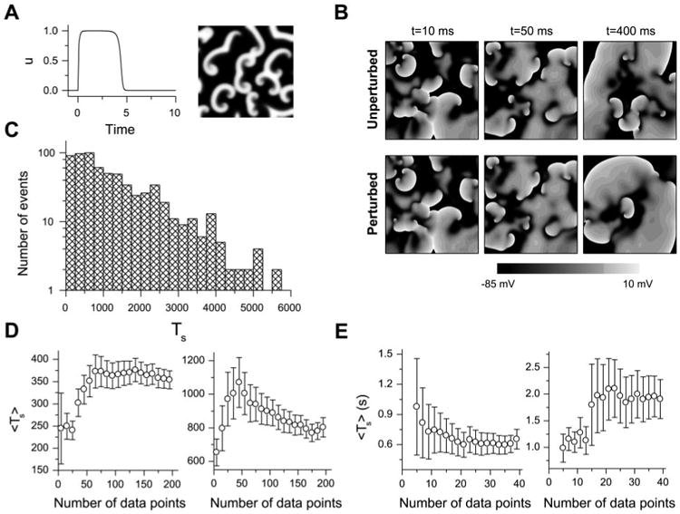
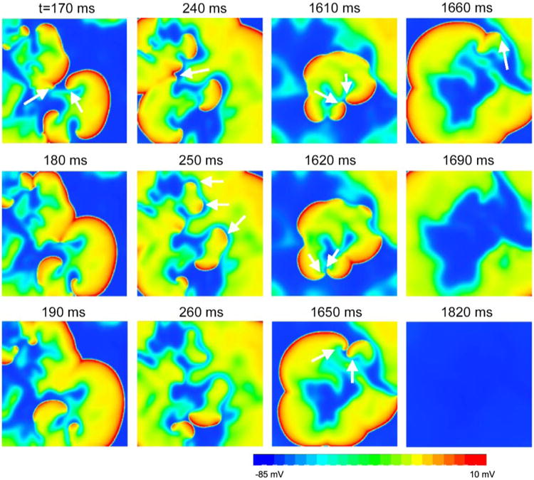
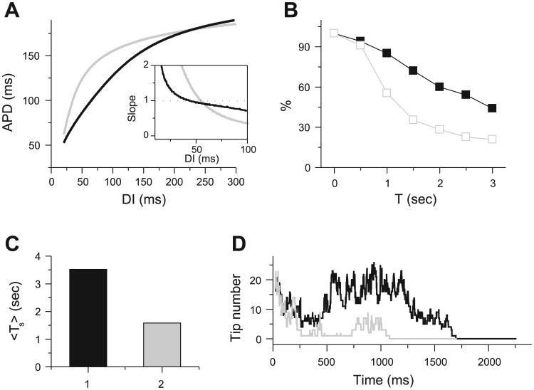
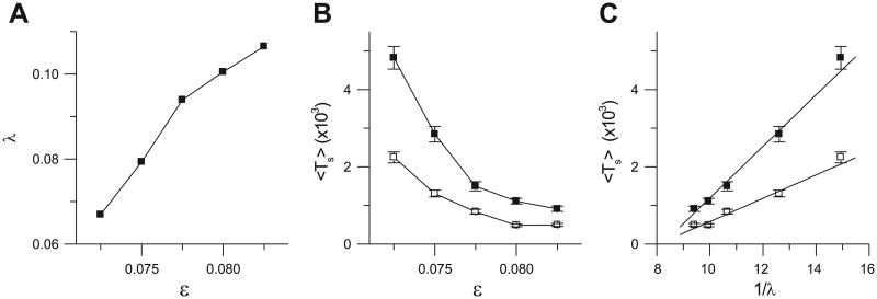
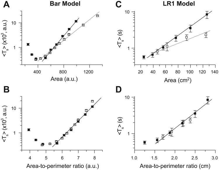
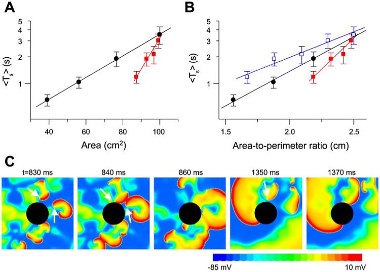
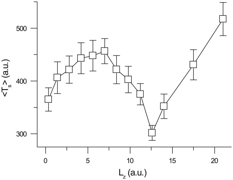
The tendency of atrial or ventricular fibrillation to terminate spontaneously in finite-sized tissue is known as the critical mass hypothesis. Previous studies have shown that dynamical instabilities play an important role in creating new wave breaks that maintain cardiac fibrillation, but its role in self-termination, in relation to tissue size and geometry, is not well understood. This study used computer simulations of two- and three-dimensional tissue models to investigate qualitatively how, in relation to tissue size and geometry, dynamical instability affects the spontaneous termination of cardiac fibrillation. The major findings are as follows: 1) Dynamical instability promotes wave breaks, maintaining fibrillation, but it also causes the waves to extinguish, facilitating spontaneous termination of fibrillation. The latter effect predominates as dynamical instability increases, so that fibrillation is more likely to self-terminate in a finite-sized tissue. 2) In two-dimensional tissue, the average duration of fibrillation increases exponentially as tissue area increases. In three-dimensional tissue, the average duration of fibrillation decreases initially as tissue thickness increases as a result of thickness-induced instability but then increases after a critical thickness is reached. Therefore, in addition to tissue mass and geometry, dynamical instability is an important factor influencing the maintenance of cardiac fibrillation.


Figures

Fig. 1
Fig. 1
A: u vs. time for a single cell (left) and a snapshot of u during spiral wave breakup in a 42 × 42 tissue (right) for the Bär model. B: comparison of simulated fibrillation patterns in a 10 × 10-cm2 tissue with the Luo and Rudy (LR1) model for an unperturbed and a randomly perturbed initial condition; at 400 ms, the patterns are very different. C: transient time (Ts) distribution in 2-dimensional (2-D) tissue with the Bär model (total 656 transients, ∊ = 0.075, tissue size = 26.25 × 26.25). D: averaged Ts (Ts) vs. number of data points used to calculate Ts for the Bär model in a 21 × 21 (left) and a 24.5 × 24.5 (right) tissue. E: Ts vs. number of data points used to calculate Ts for the LR1 model in a 7.5 × 7.5-cm2 (left) and an 8.75 × 8.75-cm2 (right) tissue.
Fig. 2
Fig. 2
Snapshots showing self-termination of “multiple-wavelet” fibrillation from a simulation in a 10 × 10-cm2 homogeneous tissue using the LR1 model. Column 1 (170–190 ms): 2 spiral waves (arrows) at 170 ms collided at 180 ms and disappeared at 190 ms. Column 2 (240–260 ms): spiral waves (arrows) at 240 and 250 ms ran into refractory tails of their previous waves and disappeared at 260 ms. Column 3 (1,610–1,650 ms): spiral pairs (arrows) collided and disappeared. Column 4 (1,660–1,820 ms): the only surviving spiral wave (arrow) moved off the tissue border, and the tissue became quiescent.
Fig. 3
Fig. 3
Effects of increasing dynamical instability on spiral wave breakup transient time in multiple-wavelet fibrillation in 10 × 10-cm2 homogeneous tissue using the LR1 model. A: 2 action potential duration (APD) restitution curves and their slopes (inset). DI, diastolic interval. Black curve: si = 0.052 mS/cm2; gray curve: si = 0.06 mS/cm2, τd → 0.75τd, τf → 0.75τf (where si is slow inward conductance and τd and τf represent activation and inactivation time constants, respectively). B: probability that Ts is longer than time T for APD restitution curves in A. C: Ts for the 2 cases in A. D: number of spiral wave tips vs. time for the 2 cases in A.
Fig. 4
Fig. 4
Relation between dynamical instability and spiral wave breakup transient time in homogeneous tissue using the Bär model. A: Lyapunov exponent (λ) vs. ∊. B: Ts vs. ∊ for 24.5 × 24.5 (□) and 26.25 × 26.25 (■) tissues. C: Ts vs. 1/λ for 24.5 × 24.5 (□) and 26.25 × 26.25 (■) tissues.
Fig. 5
Fig. 5
Effects of tissue size and geometry on Ts in homogeneous tissue. A and B: Ts vs. tissue area and area-to-perimeter ratio, respectively, for the Bär model. ■, Square tissue; □, rectangular tissue with one side fixed at 21 arbitrary units (AU). C and D: Ts vs. tissue area and area-to-perimeter ratio, respectively, for the LR1 model with the black APD restitution curve in Fig. 3A. Fixed side for rectangular tissue is 6.25 cm. [Theoretically, the function y = a + exp(αx2) should not also be the function y = b + exp(βx), but for a small range of x, a data set may well fit both functions, which may be the case here.]
Fig. 6
Fig. 6
Effects of obstacles on Ts. A: Ts vs. tissue area. ●, Replot of Ts in homogeneous square tissue of different sizes; red squares 10 × 10-cm2 tissue with a circular hole in the center (radius = 0.5, 1.0, 1.5, or 2.0 cm). B: Ts vs. area-to-outer perimeter ratio [i.e., Ts vs. (100 cm2 − πr2)/40 cm, red filled squares] and Ts vs. area-to-total perimeter ratio [i.e., Ts vs. (100 cm2 − πr2)/(40 cm + 2πr2), blue open squares] for Ts in A. ●, Replot of Ts in homogeneous square tissue vs. area-to-perimeter ratio. C: snapshots illustrating disappearance of spiral waves due to the obstacle.
Fig. 7
Fig. 7
Effects of tissue thickness (Lz) on Ts according to the Bär model with ∊ = 0.075. x- and y-dimensions were fixed as follows: Lx = Ly = 21 AU.

Similar articles

Cited by

References

    1. Allessie M, Ausma J, Schotten U. Electrical, contractile and structural remodeling during atrial fibrillation. Cardiovasc Res. 2002;54:230–246. - PubMed
    1. Allessie MA, Boyden PA, Camm AJ, Kleber AG, Lab MJ, Legato MJ, Rosen MR, Schwartz PJ, Spooner PM, Van Wagoner DR, Waldo AL. Pathophysiology and prevention of atrial fibrillation. Circulation. 2001;103:769–777. - PubMed
    1. Armoundas AA, Wu R, Juang G, Marban E, Tomaselli GF. Electrical and structural remodeling of the failing ventricle. Pharmacol Ther. 2001;92:213–230. - PubMed
    1. Bar M, Eiswirth M. Turbulence due to spiral breakup in a continuous excitable medium. Phys Rev E. 1993;48:R1635–R1637. - PubMed
    1. Calabro MP, Luzza F, Carerj S, Oreto G. Spontaneous termination of ventricular fibrillation. Heart. 2004;90:133. - PMC - PubMed

Publication types

MeSH terms