. 2005 Nov;89(5):3345-52.
doi: 10.1529/biophysj.105.068759. Epub 2005 Aug 19.

N-terminal-mediated homomultimerization of prestin, the outer hair cell motor protein

Affiliations

N-terminal-mediated homomultimerization of prestin, the outer hair cell motor protein

Dhasakumar Navaratnam et al. Biophys J. 2005 Nov.

Abstract

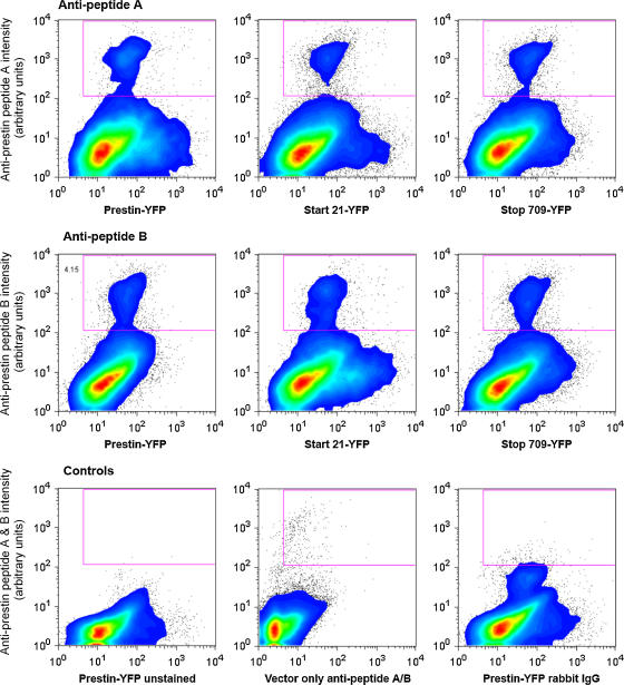
The outer hair cell lateral membrane motor, prestin, drives the cell's mechanical response that underpins mammalian cochlear amplification. Little is known about the protein's structure-function relations. Here we provide evidence that prestin is a 10-transmembrane domain protein whose membrane topology differs from that of previous models. We also present evidence that both intracellular termini of prestin are required for normal voltage sensing, with short truncations of either terminal resulting in absent or modified activity despite quantitative findings of normal membrane targeting. Finally, we show with fluorescence resonance energy transfer that prestin-prestin interactions are dependent on an intact N-terminus, suggesting that this terminus is important for homo-oligomerization of prestin. These domains, which we have perturbed, likely contribute to allosteric modulation of prestin via interactions among prestin molecules or possibly between prestin and other proteins, as well.


Figures

FIGURE 1
FIGURE 1
(A) Model of prestin's transmembrane topology that posits 10 transmembrane domains with intracellular amino and carboxy termini of the protein. Shown are the positions (red circles) of individual truncations of the protein that we tested. A cartoon of the crystal structure of the C-terminus of prestin modeled on the bacterial protein SpoIIAA, to which it shows strong homology, is shown. The positions of two peptides that would lie on the extracellular surface of the protein between transmembrane domains 4 and 5 (peptide A) and 6 and 7 (peptide B) are indicated. (B) Affinity-purified antibodies against these peptides (ApA and ApB) were used to stain both live guinea pig OHC (upper panel of confocal images) and live CHO cells transfected with prestin (lower panel). These results, together with other data (see text), confirm that these antigens lie on the extracellular surface, and support the 10-transmembrane model.
FIGURE 2
FIGURE 2
Confocal images show CHO cells transfected with prestin-YFP constructs into which the HA epitope was inserted into position 168 (AF) and position 371 (GI). The HA tags were detected with a mouse anti-HA epitope and Alexa 647-conjugated antimouse antibody (A, D, and G). (B, E, and H). YFP images. (C, F, and I) Merged images. Cells in AC and GI were stained live, and cells in D-F were fixed and permeabilized with detergent. As is evident, the detection of the HA epitope at position 168 required that the cells be permeabilized, whereas that at position 371 was detectable in live cells without permeabilization. These results suggest that position 168 of prestin in these constructs lies on the intracellular surface, whereas position 371 lies on the extracellular surface. (J) Substitution of the two potential N-glycosylation asparagine residues with glutamine residues does not result in a change in molecular weight, indicating that prestin is not glycosylated at these two residues. CHO cells were transfected with constructs of prestin fused to poly-His V5 epitope tag at its C-terminus (lane A) and prestin N163Q + N166Q double mutant fused to a poly-His V5 epitope tag at its C-terminus (lane B). Normal and mutated prestin were purified from lysed CHO cells on a Ni column and separated by PAGE (8%). The gel was blotted on to polyvinylidene difluoride and probed with an anti-V5 antibody/horseradish peroxidase-conjugated secondary antibody followed by enhanced chemiluminescence detection. As is evident, prestin migrates at its predicted molecular weight of 80 kD, as does the N163Q + N166Q double mutant.
FIGURE 3
FIGURE 3
Serial truncations of the amino and carboxy termini of prestin result in a loss of NLC. (A) Nonlinear capacitance as a function of C-terminal truncations. A stop codon at 712 abolishes NLC. (B) Examples of gating currents induced by a voltage step showing a decrease in magnitude corresponding to reductions in NLC. (C) Nonlinear capacitance as a function of N-terminal truncations. A start codon at 21 abolishes NLC. (EG) Confocal images of CHO cells transfected with prestin-YFP, start 21 prestin-YFP, and stop 709 prestin-YFP, respectively, showing plasma membrane targeting of these constructs. (H) A confocal image of a CHO cell transfected with stop 498 prestin-YFP that does not target the membrane (and corroborates work by other workers in the field; Jing Zheng, Northwestern University, and Dominik Oliver, University of Freiburg, personal communication, AOR meeting, 2004). These results confirm that the loss in NLC with these truncations is not due to absent targeting to the surface membrane.
FIGURE 4
FIGURE 4
Quantification of prestin surface expression in transfected CHO cells by flow cytometry. CHO cells transfected with prestin-YFP fusion constructs (normal prestin, start 21, and stop 709) were disassociated and stained live with two affinity-purified rabbit polyclonal antibodies to two peptides expressed on the extracellular surface of the protein (upper panels: antibody to peptide A = GGKEFNERFKEKLPAPI (aa 274–290); middle panels: antibody to peptide B = KELANKHGYQVDGNQEL (aa 359–375)). The primary antibody was detected using a biotinylated anti-rabbit antibody (Molecular Probes) and streptavidin conjugated to Alexa 647. All antibody staining was done in phosphate-buffered saline and 0.5% bovine serum albumin at 4°C. The cells were then fixed in 4% paraformaldehyde before flow cytometric analysis. Shown are contour plots with outliers of YFP intensity (Phycoerythrin window) on the x axis and prestin intensity (Alexa 647) on the y axis of live stained cells (identified by forward and side scatter). As is evident, a subpopulation of YFP positive cells, recognized by both antibodies, is present in those cells transfected with the three prestin-YFP fusion constructs (normal prestin, start 21, and stop 709) and not in those cells transfected with empty vector alone stained with anti-prestin antibodies to peptides A and B, unstained prestin-YFP transfected cells, or prestin-YFP transfected cells stained with rabbit IgG (lower panels). Both the intensity of staining and the percentage of cells within the boxed area recognized by the antibodies are similar in cells transfected with full-length prestin (4.4% and 4.3% for anti-peptides A and B, respectively), start 21 (2.6% and 3.9%), and stop 709 (3.2% and 3.3%). The three control groups contained an insignificant number of cells labeled with the antibodies. Differences in number of cells indicate variability in expression efficiency among experiments hovering around 3–6%. These efficiencies are obtained by other workers in the field (Dominik Oliver, personal communication). These quantitative analyses are evidence that the absence in NLC in these two truncations is not due to aberrant surface expression.
FIGURE 5
FIGURE 5
FRET demonstrated by acceptor (YFP) photobleaching confirms prestin-prestin interactions and suggests that the N-terminus is important in these interactions. The figure shows a CHO cell transfected with prestin-CFP (A and D) and prestin-YFP (B and E) before (AC) and after (DF) photobleaching YFP (514 line of the Argon laser) in the right half of the cell. (C and F) Merged images of A and B, and D and E, respectively. As is evident, there is an increase in CFP emission (D and F) concomitant with a decrease in YFP emission (E and F) in the right half of the cell. (G) The graph shows a quantitative estimate of FRET efficiencies after photobleaching (7). The efficiency of FRET is compared between CHO cells transfected with prestin-CFP together with prestin-YFP without (first bar) and with (second bar) acceptor photobleaching, prestin-CFP together with start 21 prestin-YFP (third bar), and prestin-CFP together with stop 709 prestin-YFP (fourth bar). A normal decrease in FRET efficiency in the absence of YFP photobleaching (−7.39 ± 2.189 (mean ± SE), n = 3 cells) is converted to an increase in FRET efficiency with photobleaching in prestin-CFP and prestin-YFP (4.64 ± 0.63, n = 5), and prestin-CFP and stop 709 prestin-YFP (4.67 ± 1.54, n = 4), confirming interactions between normal prestin molecules, and C-terminally truncated prestin molecules and normal prestin. The absence of an increase in FRET efficiency with start 21 prestin-YFP and prestin-CFP (−1.94 ± 1.57, n = 4 cells) confirms an absence of interaction between the N-terminally truncated prestin molecules and normal prestin. A one-way ANOVA between these groups demonstrated a significant difference between unbleached normal prestin and bleached normal prestin (p < 0.002) or stop 709 prestin (p < 0.002), but no difference between unbleached normal prestin and start 21 prestin. Similarly, there was a significant difference between FRET efficiencies in normal prestin and start 21 prestin (p < 0.05) transfected cells. Also noteworthy was that FRET efficiency at the membrane (6.5 ± 1.5, n = 3) was not significantly different from elsewhere in the cell.

References

    1. Zheng, J., W. Shen, D. Z. He, K. B. Long, L. D. Madison, and P. Dallos. 2000. Prestin is the motor protein of cochlear outer hair cells. Nature. 405:149–155. - PubMed
    1. Liberman, M. C., J. Gao, D. Z. He, X. Wu, S. Jia, and J. Zuo. 2002. Prestin is required for electromotility of the outer hair cell and for the cochlear amplifier. Nature. 419:300–304. - PubMed
    1. Ludwig, J., D. Oliver, G. Frank, N. Klocker, A. W. Gummer, and B. Fakler. 2001. Reciprocal electromechanical properties of rat prestin: the motor molecule from rat outer hair cells. Proc. Natl. Acad. Sci. USA. 98:4178–4183. - PMC - PubMed
    1. Meltzer, J., and J. Santos-Sacchi. 2001. Temperature dependence of non-linear capacitance in human embryonic kidney cells transfected with prestin, the outer hair cell motor protein. Neurosci. Lett. 313:141–144. - PubMed
    1. Santos-Sacchi, J., and E. Navarrete. 2002. Voltage-dependent changes in specific membrane capacitance caused by prestin, the outer hair cell lateral membrane motor. Pflugers Arch. 444:99–106. - PubMed

Publication types

MeSH terms

LinkOut - more resources