. 2005 Sep;139(1):174-85.
doi: 10.1104/pp.104.057687. Epub 2005 Aug 19.

Functional conservation of PISTILLATA activity in a pea homolog lacking the PI motif

Affiliations

Functional conservation of PISTILLATA activity in a pea homolog lacking the PI motif

Ana Berbel et al. Plant Physiol. 2005 Sep.

Abstract

Current understanding of floral development is mainly based on what we know from Arabidopsis (Arabidopsis thaliana) and Antirrhinum majus. However, we can learn more by comparing developmental mechanisms that may explain morphological differences between species. A good example comes from the analysis of genes controlling flower development in pea (Pisum sativum), a plant with more complex leaves and inflorescences than Arabidopsis and Antirrhinum, and a different floral ontogeny. The analysis of UNIFOLIATA (UNI) and STAMINA PISTILLOIDA (STP), the pea orthologs of LEAFY and UNUSUAL FLORAL ORGANS, has revealed a common link in the regulation of flower and leaf development not apparent in Arabidopsis. While the Arabidopsis genes mainly behave as key regulators of flower development, where they control the expression of B-function genes, UNI and STP also contribute to the development of the pea compound leaf. Here, we describe the characterization of P. sativum PISTILLATA (PsPI), a pea MADS-box gene homologous to B-function genes like PI and GLOBOSA (GLO), from Arabidopsis and Antirrhinum, respectively. PsPI encodes for an atypical PI-type polypeptide that lacks the highly conserved C-terminal PI motif. Nevertheless, constitutive expression of PsPI in tobacco (Nicotiana tabacum) and Arabidopsis shows that it can specifically replace the function of PI, being able to complement the strong pi-1 mutant. Accordingly, PsPI expression in pea flowers, which is dependent on STP, is identical to PI and GLO. Interestingly, PsPI is also transiently expressed in young leaves, suggesting a role of PsPI in pea leaf development, a possibility that fits with the established role of UNI and STP in the control of this process.


Figures
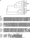
Figure 1.
Figure 1.
A, Neighbor-joining tree of B-class MADS-box deduced polypeptides from selected species: PsPI (this study; AY842491); MsNGL9 (Zucchero et al., 2001; accession no. AAK77938); AmGLO (Tröbner et al., 1992; accession no. Q03378); PhGLO1 (Angenent et al., 1992; accession no. Q03488); PhGLO2 (Kush et al., 1993; accession no. Q07474); NtGLO (Hansen et al., 1993; accession no. Q03416); AtPI (Goto and Meyerowitz, 1994; accession no. P48007); AmDEF (Sommer et al., 1990; accession no. P23706); NtDEF (Davies et al., 1996; accession no. CAA65288); PhDEF (Kush et al., 1993; accession no. CAA49567); MsNMH7 (Heard and Dunn, 1995; accession no. AAC15419); AtAP3 (Jack et al., 1992; accession no. P35632). The tree was rooted with PEAM4, a pea ortholog of AP1/SQUA (Berbel et al., 2001; accession no. AJ279089). Species names are indicated as follows: Ps, Pisum sativum; Ms, Medicago sativa; Am, Antirrhinum majus; Ph, Petunia hybrida; Nt, Nicotiana tabacum; At, Arabidopsis. B, Multiple sequence alignment of PsPI, PhGLO2, AmGLO, PhGLO1, MsNGL9, NtGLO, and AtPI. Dark gray boxes indicate fully conserved residues. Light gray boxes indicate similar and partially conserved residues.
Figure 2.
Figure 2.
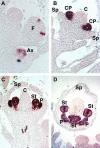
In situ hybridization of PsPI mRNA in wild-type pea inflorescences. A, Longitudinal section through a young inflorescence. PsPI expression is detected in an early stage 2 floral meristem (F) in narrow stripes of cells corresponding to the domains that will differentiate later on as common primordia of petals and stamens. PsPI is also expressed in a developing axillary bud (Ax). B, Stage 4 flower. PsPI is expressed uniformly throughout common primordia (CP) and not detected in sepal (Sp) or carpel (C) primordia. C, Stage 5 flower. The common primordia have divided to differentiate petal (P) and stamen (St) primordia that strongly express PsPI. D, Stage 7 flower. PsPI mRNA is also detected at later stages of petal and stamen development. Developmental stages were defined according to Ferrándiz et al. (1999).
Figure 3.
Figure 3.
Phenotypic effects of constitutive expression of PsPI in tobacco plants. A, Wild-type (left) and 35S::PsPI flowers (right). Mild effects of transgenic 35S::PsPI expression on sepal morphology are visible. B, Sepals from a wild-type (left) and a 35S::PsPI flower (right). 35S::PsPI sepals are longer and paler than the wild type. C to E, SEM micrographs of the adaxial side of first or second whorl organs to reveal epidermal cell morphology. C, 35S::PsPI sepal. D, Wild-type sepal. E, Wild-type petal at the basal part of the corolla tube. F to I, Phenotypic alterations in fourth whorl organ morphology found in 35S::PsPI flowers. F, Wild-type carpel. G, Fourth whorl organs of 35S::PsPI flowers with different levels of homeotic transformations. In these organs, the ovary develops abnormally, shifting toward apical positions (arrowheads). H, Wild-type stamen. I, A strongly affected 35S::PsPI carpel dissected to reveal the developing ovules. The ovary occupies an apical position in the organ, and the style is short and deformed. J to S, SEM micrographs of different cellular types. J, Epidermal cells at the basal region of a 35S::PsPI carpel. K, Epidermal cells at the basal part of the filament of a wild-type stamen, densely covered by trichomes. L, Epidermal cells at intermediate positions of a 35S::PsPI carpel. M, Epidermal cells at intermediate positions of the filament of a wild-type stamen. Trichome number gradually decreases toward the apical positions of the filament. N, Close-up of the cells in L. O, Close-up of the cells in M. P, Apical structure in a 35S::PsPI carpel. The ovary develops a central groove, resembling the morphology of an anther divided in two thecae. Q, Epidermal cells of a wild-type anther, dome-shaped and crenellated. R, Epidermal cells of the abnormal ovary of a 35S::PsPI carpel, resembling wild-type anther cell types. S, Epidermal cells of a wild-type ovary, flat-shaped and not crenellated.
Figure 4.
Figure 4.
Effects of constitutive expression of PsPI in Arabidopsis. A to H, Phenotypic effects of 35S::PsPI in Arabidopsis. A, Wild-type flower at anthesis. B, 35S::PsPI flowers at anthesis. White sectors appear on the edges of first whorl organs. Chimeric sepals are inserted at the base of the flowers at a wider angle when compared to the wild type. C, Wild-type sepals. D, 35S::PsPI first whorl organs. Patches of petal-like tissue are clearly visible. E to H, SEM micrographs of the abaxial side of first and second whorl organs. E, Epidermal cells of 35S::PsPI first whorl organs show mixed morphologies. Cells in region 1 are elongated and contain trichomes (arrow) and stomata, resembling cell morphologies of wild-type sepals shown in G. Cells in region 2 are smaller and round, similar to wild-type petal cells shown in H. F, Close-up of boxed area in E. Arrow indicates the presence of a stoma. G, Abaxial epidermal cells of a wild-type sepal. H, Abaxial epidermal cells of a wild-type petal. I to N, Phenotypic complementation of the Arabidopsis pi-1 mutant by 35S::PsPI. I, A pi-1 mutant flower, showing the typical homeotic transformations of B-class mutants. One sepal has been removed to reveal the sepal-like second whorl organs. Third whorl organs have acquired carpel identity and have incorporated into the central gynoecium. J to L, Flowers from different 35S::PsPI pi-1 lines exhibiting different degrees of phenotypic rescue. J, A partially complemented 35S::PsPI pi-1 flower. Petal formation has been fully restored, but no stamens have formed. K, A 35S::PsPI pi-1 flower in which different types of third whorl organs have developed. Two stamens have formed in lateral positions, while two carpel-like structures are visible in medial positions. L, 35S::PsPI pi-1 flower with a full complement of petals and four nearly wild-type stamens. M and N, Mosaic organs with mixed characteristics of carpel and stamens developing at the third whorl of 35S::PsPI pi-1 flowers.
Figure 5.
Figure 5.
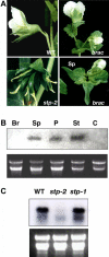
Expression of PsPI in flowers and floral organs of wild-type and pea mutant plants. A, Phenotype of pea floral mutants. Top left, wild-type (WT) flower; top right, brac mutant flower; bottom left, stp-2 mutant flower; bottom right, first whorl organs of a brac mutant flower, showing a sector of petaloid tissue. B and C, northern analysis of PsPI expression in floral organs of the brac mutant (B) or flowers of pea mutant plants (C). Blots were loaded with samples of 10 μg RNA or 15 μg RNA, respectively. As a control for equal RNA loading, a picture of each gel, stained with ethidium bromide, is shown below the blots. Br, Bract; Sp, sepal; P, petal; St, stamen; C, carpel.
Figure 6.
Figure 6.
In situ hybridization analysis of PsPI expression during axillary bud development in wild-type pea plants. A, Longitudinal section of a young vegetative pea apex. PsPI expression is detected in axillary buds from early stages of development. B, Longitudinal section through a young pea inflorescence. PsPI is strongly expressed in common primordia of the stage 4 flower at the top. Lower levels of PsPI expression are also detected in axillary buds at lower nodes. C, SEM micrograph of a developing axillary bud, showing the morphology of this structure. D to F, PsPI expression during development of the axillary bud. PsPI mRNA is detected uniformly at inception of the axillary bud but becomes progressively restricted to the developing stipules and leaves, disappearing from the axillary meristem as the bud develops. SAM, Shoot apical meristem; Ax, axillary meristem; Spl, stipule; L, leaf.

References

    1. Angenent GC, Busscher M, Franken J, Mol JNM, van Tunen AJ (1992) Differential expression of two MADS box genes in wild-type and mutant petunia flowers. Plant Cell 4: 983–993 - PMC - PubMed
    1. Bechtold N, Ellis J, Pelletier G (1993) In planta Agrobacterium mediated gene transfer by infiltration of adult Arabidopsis thaliana plants. C R Acad Sci Ser III Life Sci 316: 1194–1199
    1. Berbel A, Navarro C, Ferrándiz C, Cañas LA, Madueño F, Beltrán JP (2001) Analysis of PEAM4, the pea AP1 functional homologue, supports a model for AP1-like genes controlling both floral meristem and floral organ identity in different plant species. Plant J 25: 441–451 - PubMed
    1. Bowman JL, Smyth DR, Meyerowitz EM (1989) Genes directing flower development in Arabidopsis. Plant Cell 1: 37–52 - PMC - PubMed
    1. Carpenter R, Copsey L, Vincent C, Doyle S, Magrath R, Coen ES (1995) Control of flower development and phyllotaxy by meristem identity genes in Antirrhinum. Plant Cell 7: 2001–2011 - PMC - PubMed

Publication types

MeSH terms