Incomplete activation of macrophage apoptosis during intracellular replication of Legionella pneumophila
- PMID: 16113249
- PMCID: PMC1231138
- DOI: 10.1128/IAI.73.9.5339-5349.2005
Incomplete activation of macrophage apoptosis during intracellular replication of Legionella pneumophila
Abstract
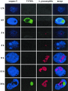
The ability of the intracellular bacterium Legionella pneumophila to cause disease is totally dependent on its ability to modulate the biogenesis of its phagosome and to replicate within alveolar cells. Upon invasion, L. pneumophila activates caspase-3 in macrophages, monocytes, and alveolar epithelial cells in a Dot/Icm-dependent manner that is independent of the extrinsic or intrinsic pathway of apoptosis, suggesting a novel mechanism of caspase-3 activation by this intracellular pathogen. We have shown that the inhibition of caspase-3 prior to infection results in altered biogenesis of the L. pneumophila-containing phagosome and in an inhibition of intracellular replication. In this report, we show that the preactivation of caspase-3 prior to infection does not rescue the intracellular replication of L. pneumophila icmS, icmR, and icmQ mutant strains. Interestingly, preactivation of caspase-3 through the intrinsic and extrinsic pathways of apoptosis in both human and mouse macrophages inhibits intracellular replication of the parental stain of L. pneumophila. Using single-cell analysis, we show that intracellular L. pneumophila induces a robust activation of caspase-3 during exponential replication. Surprisingly, despite this robust activation of caspase-3 in the infected cell, the host cell does not undergo apoptosis until late stages of infection. In sharp contrast, the activation of caspase-3 by apoptosis-inducing agents occurs concomitantly with the apoptotic death of all cells that exhibit caspase-3 activation. It is only at a later stage of infection, and concomitant with the termination of intracellular replication, that the L. pneumophila-infected cells undergo apoptotic death. We conclude that although a robust activation of caspase-3 is exhibited throughout the exponential intracellular replication of L. pneumophila, apoptotic cell death is not executed until late stages of the infection, concomitant with the termination of intracellular replication.
Figures


Similar articles
-
Activation of caspase-3 by the Dot/Icm virulence system is essential for arrested biogenesis of the Legionella-containing phagosome.Cell Microbiol. 2004 Jan;6(1):33-48. doi: 10.1046/j.1462-5822.2003.00335.x. Cell Microbiol. 2004. PMID: 14678329
-
Anti-apoptotic signalling by the Dot/Icm secretion system of L. pneumophila.Cell Microbiol. 2007 Jan;9(1):246-64. doi: 10.1111/j.1462-5822.2006.00785.x. Epub 2006 Aug 15. Cell Microbiol. 2007. PMID: 16911566
-
Host-dependent trigger of caspases and apoptosis by Legionella pneumophila.Infect Immun. 2007 Jun;75(6):2903-13. doi: 10.1128/IAI.00147-07. Epub 2007 Apr 9. Infect Immun. 2007. PMID: 17420236 Free PMC article.
-
Molecular and cell biology of Legionella pneumophila.Int J Med Microbiol. 2004 Apr;293(7-8):519-27. doi: 10.1078/1438-4221-00286. Int J Med Microbiol. 2004. PMID: 15149027 Review.
-
Modulation of caspases and their non-apoptotic functions by Legionella pneumophila.Cell Microbiol. 2010 Feb;12(2):140-7. doi: 10.1111/j.1462-5822.2009.01401.x. Epub 2009 Oct 27. Cell Microbiol. 2010. PMID: 19863553 Review.
Cited by
-
Legionella pneumophila glucosyltransferase inhibits host elongation factor 1A.Proc Natl Acad Sci U S A. 2006 Nov 7;103(45):16953-8. doi: 10.1073/pnas.0601562103. Epub 2006 Oct 26. Proc Natl Acad Sci U S A. 2006. PMID: 17068130 Free PMC article.
-
Phagocytosis of Staphylococcus aureus by macrophages exerts cytoprotective effects manifested by the upregulation of antiapoptotic factors.PLoS One. 2009;4(4):e5210. doi: 10.1371/journal.pone.0005210. Epub 2009 Apr 21. PLoS One. 2009. PMID: 19381294 Free PMC article.
-
Complete and ubiquitinated proteome of the Legionella-containing vacuole within human macrophages.J Proteome Res. 2015 Jan 2;14(1):236-48. doi: 10.1021/pr500765x. Epub 2014 Nov 13. J Proteome Res. 2015. PMID: 25369898 Free PMC article.
-
Asc-dependent and independent mechanisms contribute to restriction of legionella pneumophila infection in murine macrophages.Front Microbiol. 2011 Feb 14;2:18. doi: 10.3389/fmicb.2011.00018. eCollection 2011. Front Microbiol. 2011. PMID: 21713115 Free PMC article.
-
Temporal and spatial trigger of post-exponential virulence-associated regulatory cascades by Legionella pneumophila after bacterial escape into the host cell cytosol.Environ Microbiol. 2010 Mar;12(3):704-15. doi: 10.1111/j.1462-2920.2009.02114.x. Epub 2009 Dec 2. Environ Microbiol. 2010. PMID: 19958381 Free PMC article.
References
-
- Abu Kwaik, Y. 1998. Fatal attraction of mammalian cells to Legionella pneumophila. Mol. Microbiol. 30:689-696. - PubMed
-
- Algeciras-Schimnich, A., B. C. Barnhart, and M. E. Peter. 2002. Apoptosis-independent functions of killer caspases. Curr. Opin. Cell Biol. 14:721-726. - PubMed
-
- Baud, V., and M. Karin. 2001. Signal transduction by tumor necrosis factor and its relatives. Trends Cell Biol. 11:372-377. - PubMed
-
- Berger, K. H., J. Merriam, and R. R. Isberg. 1994. Altered intracellular targeting properties associated with mutations in the Legionella pneumophila dotA gene. Mol. Microbiol. 14:809-822. - PubMed
-
- Bertrand, R., E. Solary, P. O'Connor, K. W. Kohn, and Y. Pommier. 1994. Induction of a common pathway of apoptosis by staurosporine. Exp. Cell Res. 211:314-321. - PubMed
Publication types
MeSH terms
Substances
Grants and funding
LinkOut - more resources
Full Text Sources
Other Literature Sources
Research Materials

