. 2005 Sep;73(9):5379-87.
doi: 10.1128/IAI.73.9.5379-5387.2005.

Gliding motility leads to active cellular invasion by Cryptosporidium parvum sporozoites

Affiliations

Gliding motility leads to active cellular invasion by Cryptosporidium parvum sporozoites

Dawn M Wetzel et al. Infect Immun. 2005 Sep.

Abstract

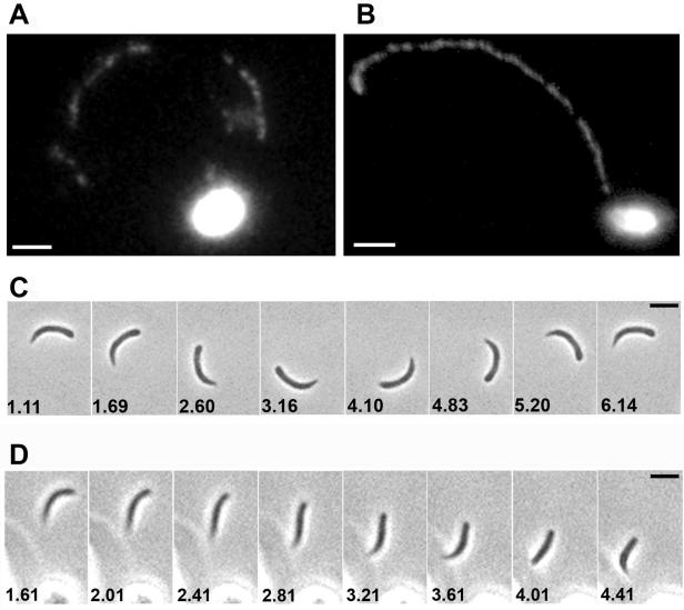
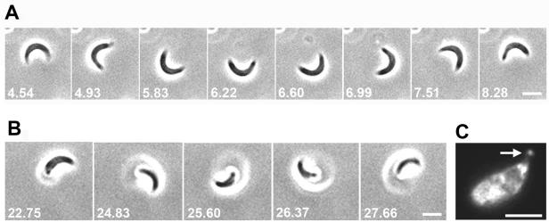
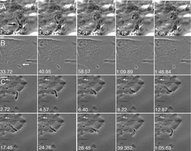
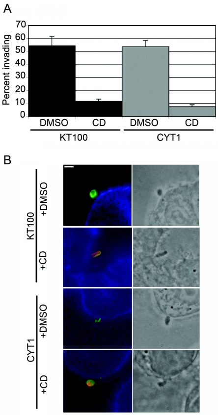
We examined gliding motility and cell invasion by an early-branching apicomplexan, Cryptosporidium parvum, which causes diarrheal disease in humans and animals. Real-time video microscopy demonstrated that C. parvum sporozoites undergo circular and helical gliding, two of the three stereotypical movements exhibited by Toxoplasma gondii tachyzoites. C. parvum sporozoites moved more rapidly than T. gondii sporozoites, which showed the same rates of motility as tachyzoites. Motility by C. parvum sporozoites was prevented by latrunculin B and cytochalasin D, drugs that depolymerize the parasite actin cytoskeleton, and by the myosin inhibitor 2,3-butanedione monoxime. Imaging of the initial events in cell entry by Cryptosporidium revealed that invasion occurs rapidly; however, the parasite does not enter deep into the cytosol but rather remains at the cell surface in a membrane-bound compartment. Invasion did not stimulate rearrangement of the host cell cytoskeleton and was inhibited by cytochalasin D, even in host cells that were resistant to the drug. Our studies demonstrate that C. parvum relies on a conserved actin-myosin motor for motility and active penetration of its host cell, thus establishing that this is a widely conserved feature of the Apicomplexa.


Figures

FIG. 1.
FIG. 1.
Gliding motility by C. parvum sporozoites. (A and B) C. parvum deposited surface membrane trails during gliding on a solid substrate. (Panels A and B show two separate examples.) Trails were visualized by immunofluorescence microscopy using an antibody to the surface protein p25 (mouse monoclonal antibody 3E3) and a secondary anti-mouse antibody conjugated to Oregon Green. Bar, 5 μm. (C and D) Time lapse phase-contrast video microscopy allowed visualization of gliding motility by C. parvum in real time. Shown are series of frames from movies of circular gliding (video 1) (C) and helical gliding (video 2) (D) by C. parvum sporozoites. Time elapsed is shown in seconds in the lower left corner of each frame. Bars, 5 μm.
FIG. 2.
FIG. 2.
Gliding motility by T. gondii sporozoites. (A and B) Time lapse phase-contrast video microscopy allowed visualization of gliding motility by T. gondii sporozoites in real time. Shown are series of frames from movies of circular gliding (video 3) (A) and twirling (video 5) (B) by T. gondii sporozoites. Time elapsed is shown in seconds in the lower left corner of each frame. Bars, 5 μm. (C) Upon treatment with 2 μM JAS, T. gondii sporozoites formed apical projections of actin (arrow). Actin was visualized using a monoclonal antibody to actin (C4) and a secondary anti-mouse antibody conjugated to Texas Red. Bar, 5 μm.
FIG. 3.
FIG. 3.
Real-time invasion of host cells by C. parvum. (A) C. parvum invading a KB100 epithelial cell (video 6). The parasite was gliding on top of the cell monolayer prior to invasion. (B) C. parvum invading an MDCK cell (video 7). The parasite approached the edge of the cell and then invaded just under the cell membrane. (C) C. parvum appearing to enter and exit a KB100 cell (video 8). Bar, 5 μm. All images are from video 3. Arrows indicate the position of the parasite in the first frame.
FIG. 4.
FIG. 4.
C. parvum invasion depends on parasite actin but not on host cell actin. (A) C. parvum efficiently invaded KB and CYT1 cells in the absence of drug (DMSO control). In the presence of CD, the percentage of parasites invading KB100 cells was reduced from 54 to 11% (P < 0.05), and the percentage of parasites invading CYT1 cells, which are resistant to CD, was reduced from 53 to 7% (P < 0.05). The percentage invading was calculated using a two-color latency assay described in the legend to panel B. Results shown are means ± SE (n = 3). (B) A two-color immunofluorescence assay demonstrated that C. parvum was able to efficiently invade host cells yet remained at the cell surface and often protruded away from the cell surface despite being enveloped within the host cell membrane (DMSO control). Cryptosporidium was not able to invade either control or CD-resistant host cells in the presence of CD. Parasites were allowed to enter KB100 or CYT1 cells, fixed, and stained by using the antibody to surface p25 followed by secondary antibodies conjugated to Alexa 594 (red). Cells were then permeabilized and stained with anti-p25 followed by secondary antibodies conjugated to Alexa 488 (green). External parasites were labeled red and green (and appear orange-yellow in most cases), and parasites that had invaded cells were labeled green. Host cell actin was labeled with phalloidin conjugated to Alexa 350 (blue). Shown are merged 3-color immunofluorescence images (left) and the corresponding phase images (right). Bar, 5 μm.

References

    1. Allen, M. L., J. M. Dobrowolski, H. Muller, L. D. Sibley, and T. E. Mansour. 1997. Cloning and characterization of actin depolymerizing factor from Toxoplasma gondii. Mol. Biochem. Parasitol. 88:43-52. - PubMed
    1. Arrowood, M. J., C. R. Sterling, and M. C. Healey. 1991. Immunofluorescent microscopical visualization of trails left by gliding Cryptosporidium parvum sporozoites. J. Parasitol. 77:315-317. - PubMed
    1. Barragan, A., F. Brossier, and L. D. Sibley. 2005. Transepithelial migration of Toxoplasma gondii involves an interaction of intercellular adhesion molecule 1 (ICAM-1) with the parasite adhesin MIC2. Cell. Microbiol. 7:561-568. - PubMed
    1. Barragan, A., and L. D. Sibley. 2003. Migration of Toxoplasma gondii across biological barriers. Trends Microbiol. 11:426-430. - PubMed
    1. Barragan, A., and L. D. Sibley. 2002. Transepithelial migration of Toxoplasma gondii is linked to parasite motility and virulence. J. Exp. Med. 195:1625-1633. - PMC - PubMed

Publication types

LinkOut - more resources