. 2005 Sep;73(9):5504-13.
doi: 10.1128/IAI.73.9.5504-5513.2005.

Mannheimia haemolytica leukotoxin induces apoptosis of bovine lymphoblastoid cells (BL-3) via a caspase-9-dependent mitochondrial pathway

Affiliations

Mannheimia haemolytica leukotoxin induces apoptosis of bovine lymphoblastoid cells (BL-3) via a caspase-9-dependent mitochondrial pathway

Dhammika N Atapattu et al. Infect Immun. 2005 Sep.

Abstract

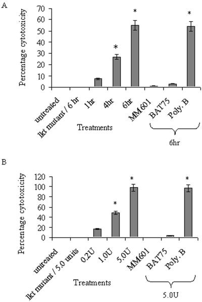
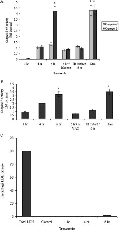
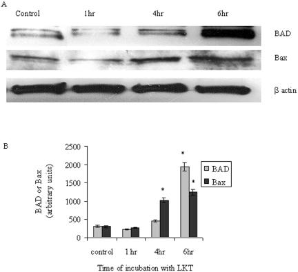
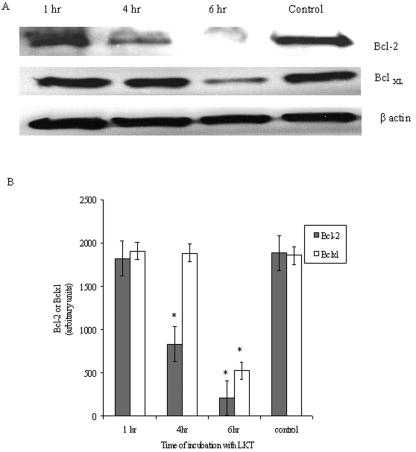
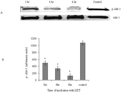
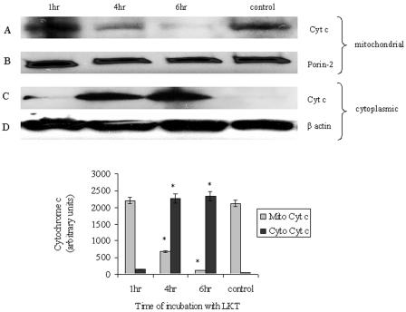
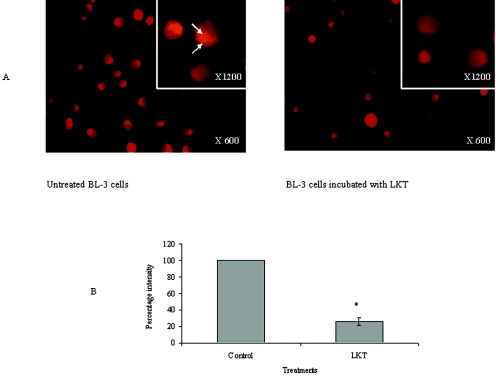
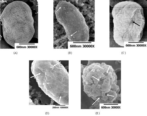
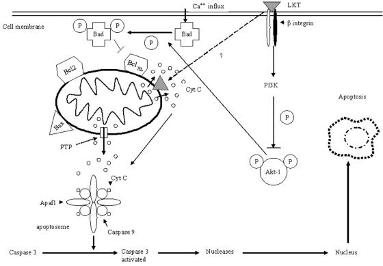
Mannheimia haemolytica is a key pathogen in the bovine respiratory disease complex. It produces a leukotoxin (LKT) that is an important virulence factor, causing cell death in bovine leukocytes. The LKT binds to the beta(2) integrin CD11a/CD18, which usually activates signaling pathways that facilitate cell survival. In this study, we investigated mechanisms by which LKT induces death in bovine lymphoblastoid cells (BL-3). Incubation of BL-3 cells with a low concentration of LKT results in the activation of caspase-3 and caspase-9 but not caspase-8. Similarly, the proapoptotic proteins Bax and BAD were significantly elevated, while the antiapoptotic proteins Bcl-2, Bcl(XL) and Akt-1 were downregulated. Following exposure to LKT, we also observed a reduction in mitochondrial cytochrome c and corresponding elevation of cytosolic cytochrome c, suggesting translocation from the mitochondrial compartment to the cytosol. Consistent with this observation, tetramethylrhodamine ethyl ester perchlorate staining revealed that mitochondrial membrane potential was significantly reduced. These data suggest that LKT induces apoptosis of BL-3 cells via a caspase-9-dependent mitochondrial pathway. Furthermore, scanning electron micrographs of mitochondria from LKT-treated BL-3 cells revealed lesions in the outer mitochondrial membrane, which are larger than previous reports of the permeability transition pore through which cytochrome c is usually released.


Figures

FIG. 1.
FIG. 1.
Cytotoxicity of M. haemolytica LKT for BL-3 cells. In panel A, 106 BL-3 cells were incubated at 37°C with 0.2 U of M. haemolytica LKT or LKT prepared from the lktC gene mutant for the designated time periods. Cells were washed three times with RPMI, and viability was assessed as described in Materials and Methods. LKT preincubated with 5 μl of anti-LKT MAb (MM601) or preincubating BL-3 cells with 5 μl anti-CD11a/CD18 (BAT75) blocked LKT-mediated cytotoxicity. LKT pretreated with 10 μg/ml polymyxin B (Poly-B) did not diminish cytotoxicity, suggesting that LPS was not responsible for cell death. The inactive LKT produced by the lktC gene mutant did not cause any cytotoxicity when incubated with BL-3 cells for 6 h at 37°C. (B) BL-3 cells were incubated for 6 h with the indicated amounts of LKT. The figure illustrates the mean ± standard error of the mean (SEM) for five separate experiments (*, P < 0.05).
FIG. 2.
FIG. 2.
M. haemolytica LKT activates caspase-3 and caspase-9 but does not increase caspase-8 activity or LDH release in BL-3 cells. BL-3 (106) cells were incubated at 37°C with LKT (0.2 U) or LKT prepared from lktC gene mutant for the designated time periods. The cells were washed three times with RPMI and lysed, and caspase-8 and caspase-9 (A) and caspase-3 (B) activity assays were performed. Some cultures of BL-3 cells were preincubated with 50 μM of a caspase-8 inhibitor (Z-EITD-fmk), caspase-9 inhibitor (Z-LEHD-fmk), or a caspase-3 and caspase-7 inhibitor (Z-VAD-fmk) for 1 h at 37°C before the addition of LKT for 6 h. Incubating BL-3 cells with 0.3 μM staurosporine for 1 h at 37°C served as a positive control for apoptosis. (C) LDH release assay showed no significant release of LDH in BL-3 cells treated with 0.2 U of LKT for the designated time periods. The data illustrate the mean ± SEM for five separate experiments (*, P < 0.05).
FIG. 3.
FIG. 3.
M. haemolytica LKT upregulates the proapoptotic proteins BAD and Bax. BL-3 cells (106) were incubated with LKT (0.2 U) for the designated time periods at 37°C. Cells were washed three times with RPMI and lysed with Triton-X lysis buffer at 4°C for 20 min, and Western immunoblotting was performed for BAD, Bax, and β-actin. (B) Images shown were quantified with a densitometer. The data are presented as means ± SEM of three separate experiments (*, P < 0.05).
FIG. 4.
FIG. 4.
M. haemolytica LKT downregulates the antiapoptotic proteins Bcl-2 and BclXL. BL-3 cells were incubated with LKT (0.2 U) for the designated time periods at 37°C. Cells were washed three times with RPMI, lysed with Triton-X lysis buffer at 4°C for 20 min, and Western immunoblotting was performed for Bcl-2, BclXL, and β-actin. (B) Images shown were quantified with a densitometer; the data are illustrated as means ± SEM of three separate experiments (*, P < 0.05).
FIG. 5.
FIG. 5.
M. haemolytica LKT downregulates p-Akt-1 in BL-3 cells. BL-3 cells (106) were treated with 0.2 U of LKT for the designated time periods at 37°C. Cells were washed three times with RPMI and lysed with Triton-X lysis buffer at 4°C for 20 min, and Western immunoblotting was performed for p-Akt-1. Blots were stripped and reprobed for Akt-1. (B) Images shown were quantified with a densitometer, and the data are illustrated as means ± SEM of three separate experiments (*, P < 0.05).
FIG. 6.
FIG. 6.
Incubation of BL-3 cells with M. haemolytica LKT results in mitochondrial release of cytochrome c. BL-3 cells (106) were incubated with of LKT (0.2 U) at 37°C for the designated time periods. Cells were washed three times with RPMI and lysed with mitochondrial lysis buffer at 4°C for 20 min, and the mitochondrial pellet and cytosolic proteins were used separately for Western immunoblotting for cytochrome c. Blots were stripped and reprobed for the mitochondrial marker protein porin and the cytosolic protein β-actin. The latter was not observed in the mitochondrial extracts (data not shown). The top two rows of blots represent mitochondrial extracts and the bottom row of blots represents cytosolic extracts from LKT-treated BL-3 cells. In the bottom panel, images shown were quantified with a densitometer, and the data are illustrated as means ± SEM of three separate experiments (*, P < 0.05).
FIG. 7.
FIG. 7.
M. haemolytica LKT diminishes mitochondrial membrane potential (Δψm). BL-3 cells (106) were treated with LKT (0.2 U) for 6 h at 37°C. Cells were washed with RPMI, stained with 0.5 mM TMRE for 10 min, and washed three times with PBS. (A) Fluorescent microscopy was performed at an emission wavelength of 575 nm (magnification, ×600). The insert panels show images of individual cells stained with TMRE at a magnification of ×1,200. Untreated control BL-3 cells show staining of mitochondria as orange-red specks distributed in the cytoplasm (arrows). BL-3 cells incubated with LKT show a significant reduction in staining of the mitochondrial matrices, due to the loss of mitochondrial membrane potential. Arrows indicate individual mitochondria stained with TMRE. (B) Images were quantified using the Image J system and statistically analyzed. The graph represents means ± SEM fluorescent intensities of 200 cells, randomly counted, in each of three separate experiments (*, P < 0.05).
FIG. 8.
FIG. 8.
Scanning electron microscopy of mitochondria isolated from BL-3 cells incubated with M. haemolytica LKT shows morphological evidence of OMM damage. Scanning electron micrographs of mitochondria isolated from control BL-3 cells (A) or BL-3 cells incubated with 0.2 U LKT for 6 h (B). (C to E) Arrows indicate blebs on the surface (C), furrows and ridges with gross deformities of the outer mitochondrial membrane (D), and large perforations and punched-out lesions (E) on mitochondria from LKT-treated BL-3 cells.
FIG. 9.
FIG. 9.
Schematic representation of mechanisms by which M. haemolytica LKT may induce apoptotic cell death in BL-3 cells.

References

    1. Adrain, C., E. M. Creagh, and S. J. Martin. 2001. Apoptosis-associated release of Smac/DIABLO from mitochondria requires active caspases and is blocked by Bcl-2. EMBO J. 20:6627-6636. - PMC - PubMed
    1. Ambagala, T. C., A. P. Ambagala, and S. Srikumaran. 1999. The leukotoxin of Pasteurella haemolytica binds to beta(2) integrins on bovine leukocytes. FEMS Microbiol Lett. 179:161-167. - PubMed
    1. Antonsson, B. 2004. Mitochondria and the Bcl-2 family proteins in apoptosis signaling pathways. Mol. Cell. Biochem. 256-257:141-155. - PubMed
    1. Araya, R., T. Uehara, and Y. Nomura. 1998. Hypoxia induces apoptosis in human neuroblastoma SK-N-MC cells by caspase activation accompanying cytochrome c release from mitochondria. FEBS Lett. 439:168-172. - PubMed
    1. Aronis, A., J. A. Melendez, O. Golan, S. Shilo, N. Dicter, and O. Tirosh. 2003. Potentiation of Fas-mediated apoptosis by attenuated production of mitochondria-derived reactive oxygen species. Cell Death Differ. 10:335-344. - PubMed

Publication types