

Experimental infection model for Johne's disease in sheep
- PMID: 16113277
- PMCID: PMC1231139
- DOI: 10.1128/IAI.73.9.5603-5611.2005
Experimental infection model for Johne's disease in sheep
Abstract
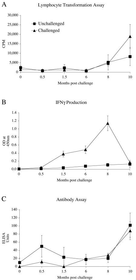
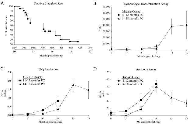
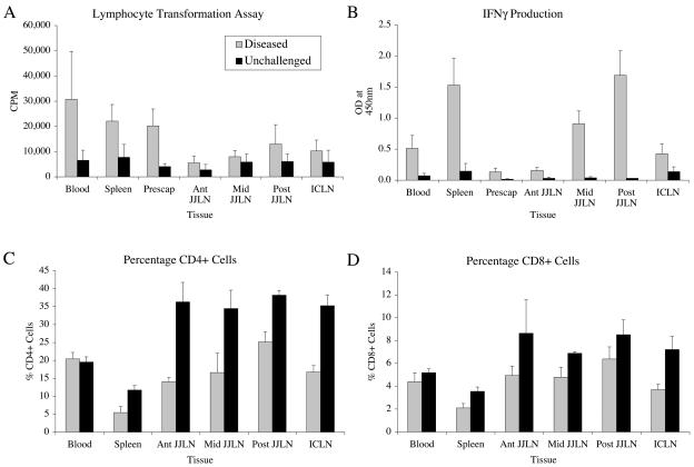
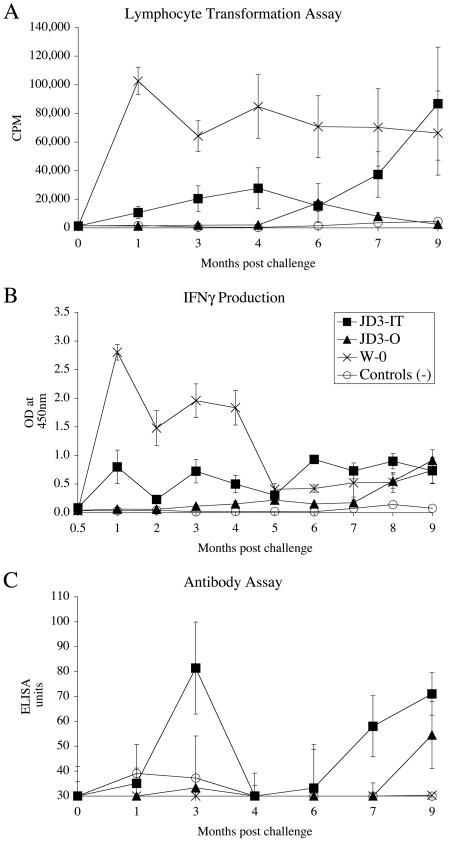
Johne's disease in ruminants results in chronic enteritis caused by the pathogenic bacterium Mycobacterium avium subsp. paratuberculosis. This study examined two M. avium subsp. paratuberculosis strains (JD3 and W), using different doses and routes of infection, to establish the optimal time postchallenge when predictable levels of infection, gut lesions, and clinical disease occur in a large proportion of sheep. While a small proportion (25%) of sheep challenged with a low-passage-number laboratory culture of M. avium subsp. paratuberculosis (strain W) became infected, no infection was found in animals exposed to a high-passage-number culture isolate of strain W. In contrast, a primary tissue homogenate of M. avium subsp. paratuberculosis (JD3) resulted in high (90%) infection rates and gut histopathology following oral or intratonsillar challenge. The optimal conditions necessary to produce Johne's disease involve oral inoculation of 3-month-old lambs with four doses of 5 x 10(8) CFU of M. avium subsp. paratuberculosis isolated directly from the gut lymphatic tissues of clinically affected sheep. This resulted in consistent gut histopathology at 9 months and the onset of clinical disease by 11 months postchallenge.
Figures
References
-
- Armstrong, M. C. 1956. Johne's disease of sheep in the South Island of New Zealand. N. Z. Vet. J. 4:56-59.
-
- Bakker, D., P. T. Willemsen, and F. G. van Zijderveld. 2000. Paratuberculosis recognized as a problem at last: a review. Vet. Q. 22:200-204. - PubMed
-
- Begara-McGorum, I., L. A. Wildblood, C. J. Clarke, K. M. Connor, K. Stevenson, C. J. McInnes, J. M. Sharp, and D. G. Jones. 1998. Early immunopathological events in experimental ovine paratuberculosis. Vet. Immunol. Immunopathol. 63:265-287. - PubMed
-
- Begara-McGorum, I. M., L. A. Wildblood, and D. G. Jones. 1997. Early immune events following experimental infection of lambs with Mycobacterium avium subspecies paratuberculosis. Biochem. Soc. Trans. 25:279S. - PubMed
-
- Billman-Jacobe, H., M. Carrigan, F. Cockram, L. A. Corner, I. J. Gill, J. F. Hill, T. Jessep, A. R. Milner, and P. R. Wood. 1992. A comparison of the interferon gamma assay with the absorbed ELISA for the diagnosis of Johne's disease in cattle. Aust. Vet. J. 69:25-28. - PubMed
Publication types
MeSH terms
LinkOut - more resources
Full Text Sources
