. 2005 Sep;73(9):5809-16.
doi: 10.1128/IAI.73.9.5809-5816.2005.

Tracking antigen-specific CD8 T lymphocytes in the lungs of mice vaccinated with the Mtb72F polyprotein

Affiliations

Tracking antigen-specific CD8 T lymphocytes in the lungs of mice vaccinated with the Mtb72F polyprotein

Scott M Irwin et al. Infect Immun. 2005 Sep.

Abstract

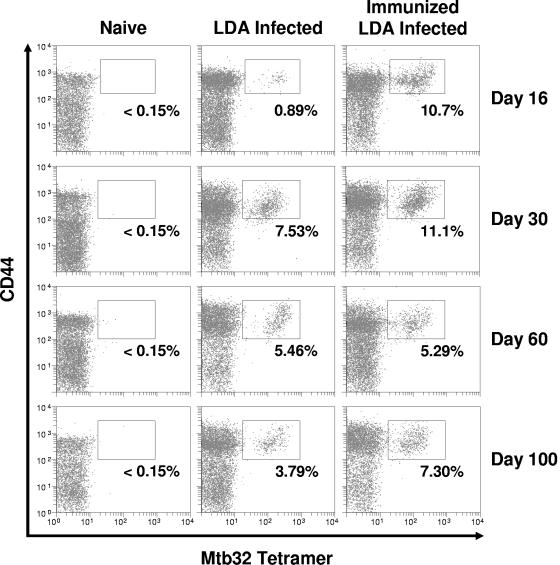
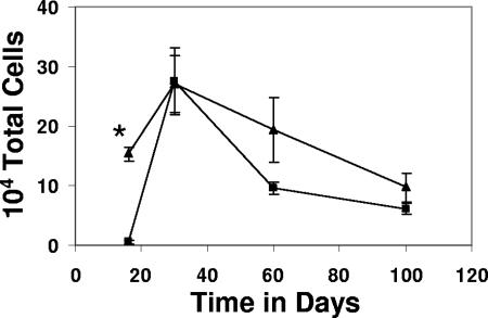
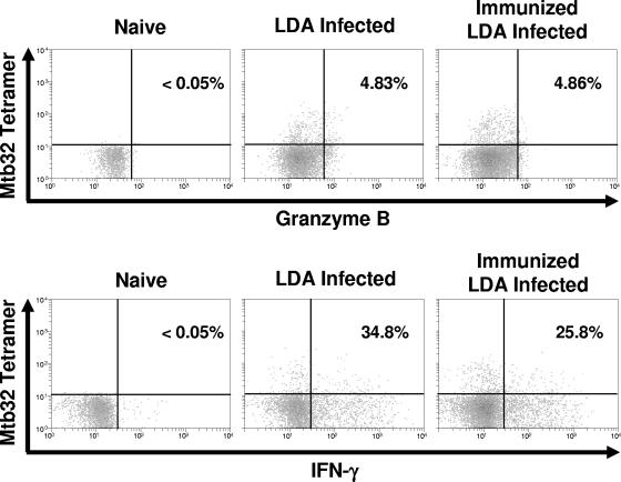
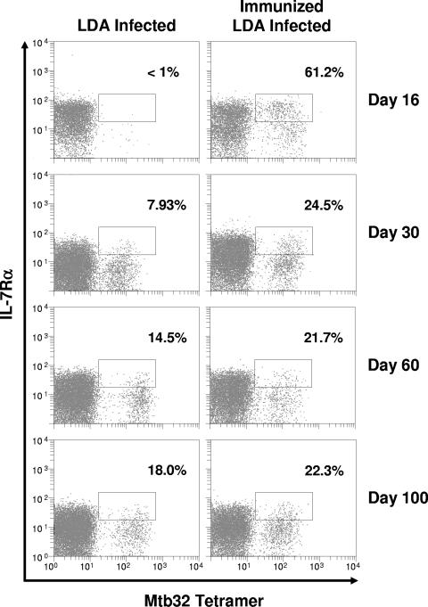
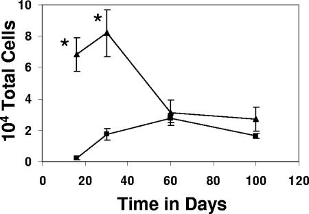
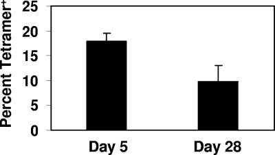
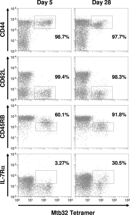
This study used a major histocompatibility complex class I tetramer reagent to track antigen-specific CD8 T cells in the lungs of mice immunized with the tuberculosis vaccine candidate Mtb72F. The results show that CD8 T cells recognizing an immunodominant Mtb32-specific epitope could be detected in significant numbers over the course of infection in mice exposed to low-dose aerosol challenge with Mycobacterium tuberculosis and that prior vaccination substantially increased the numbers of these cells early in the lungs. The effector phenotype of the cells was shown by the demonstration that many secreted gamma interferon, but very few contained granzyme B. As the course of the infection progressed, many activated CD8 T cells down-regulated expression of CD45RB and upregulated expression of the interleukin-7 receptor alpha chain, indicating a transition of these cells to a state of memory. These data support the hypothesis that M. tuberculosis-specific CD8 T cells can be targeted by vaccination with the Mtb72F polyprotein.


Figures

FIG. 1.
FIG. 1.
Kinetic analysis of the Mtb32 antigen-specific CD8-T-cell response in nonimmunized mice and in mice immunized with Mtb72F followed by low-dose aerosol (LDA) infection with M. tuberculosis. C57BL/6 mice were immunized three times, at 3-week intervals, via the intramuscular route. Three weeks following the last immunization, mice were challenged. Time points represent the number of days after infection. Lung cells were stained with the Mtb32 H-2Db tetramer and antibodies specific for CD3, CD8, and CD44. Lymphocytes were gated based upon characteristic size and granularity and further gated based upon concurrent CD3 and CD8 expression. The data are from one mouse (representative of n = 4 mice). Percentages represent the number of CD44hi tetramer-positive cells as compared to the total number of CD3/CD8-positive cells. In all cases, background tetramer staining in naïve mice was <0.15%.
FIG. 2.
FIG. 2.
Immunization with Mtb72F resulted in greater numbers of antigen-specific CD8 T cells in the lungs of mice following infection. Data are expressed as the means ± the standard errors of the means and are from four mice per group per time point. Triangles, Mtb72F-immunized mice; squares, nonimmunized mice. Statistical significance based upon a comparison of Mtb72F-immunized and nonimmunized groups was determined using Student's t test and is indicated by an asterisk (P < 0.005).
FIG. 3.
FIG. 3.
Intracellular cytokine staining of lung-derived cells for granzyme B and IFN-γ at 30 days following aerogenic infection suggests that the tetramer-positive cells are primarily IFN-γ producing rather than cytolytic. The data are from one mouse (representative of n = 4 mice). Percentages represent the number of antigen-specific IFN-γ-positive or granzyme B-positive cells as compared to the total number of tetramer-positive cells.
FIG. 4.
FIG. 4.
Mtb32 tetramer-positive CD8 T cells in the lung express high levels of IL-7Rα following immunization with Mtb72F and subsequent aerogenic challenge. At multiple time points, lung cells were stained with the H-2Db tetramer and antibodies specific for CD3, CD8, and IL-7Rα. Lymphocytes were gated based upon characteristic size and granularity and further gated based upon concurrent CD3 and CD8 expression. The data are from one mouse (representative of n = 4 mice). Percentages represent the number of IL-7Rαhi tetramer-positive cells as compared to the total number of tetramer-positive cells. In all cases, background tetramer staining in naïve mice was <0.15%.
FIG. 5.
FIG. 5.
Greater numbers of memory precursor cells were present in the lungs of Mtb72F-immunized mice prior to day 60 as compared to nonimmunized mice following M. tuberculosis infection. Data are expressed as the means ± the standard errors of the means (n = 4 mice/group/time point). Triangles, Mtb72F-immunized mice; squares, nonimmunized mice. Statistical significance based upon a comparison of Mtb72F-immunized and nonimmunized groups was determined using Student's t test and is indicated by asterisks (P < 0.05).
FIG. 6.
FIG. 6.
Immunization with Mtb72F protein in conjunction with cationic liposome complexes and noncoding DNA results in a strong pulmonary antigen-specific CD8-T-cell response in the absence of infection. C57BL/6 mice (n = 4 mice/group/time point) were immunized two times, 14 days apart, via the intraperitoneal route. Five days and 28 days following the second immunization, lung cells were isolated and stained with the H-2Db tetramer and antibodies specific for CD3, CD8, and CD44. Lymphocytes were gated based upon characteristic size and granularity and further gated based upon concurrent CD3 and CD8 expression. Data are expressed as the means ± the standard errors of the means.
FIG. 7.
FIG. 7.
Mtb32-specific CD8 T cells isolated from lungs of mice following immunization possess an activated phenotype. Mice (n = 4 mice/group/time point) were immunized two times, 14 days apart via the intraperitoneal route with cationic liposomes plus Mtb72F protein. Five days and 28 days following the second immunization, lung cells were isolated and stained with the H-2Db tetramer and antibodies specific for CD3, CD8, and surface activation markers. Lymphocytes were gated based upon characteristic size and granularity and further gated based upon concurrent CD3 and CD8 expression. Representative dot plots are shown where the percentages represent the number of tetramer-positive cells falling within the indicated region as compared to the total number of tetramer-positive cells. In all cases, background tetramer staining in naïve mice was <0.15%.

References

    1. Andersen, P. 2001. TB vaccines: progress and problems. Trends Immunol. 22:160-168. - PubMed
    1. Behar, S. M., C. C. Dascher, M. J. Grusby, C. R. Wang, and M. B. Brenner. 1999. Susceptibility of mice deficient in CD1D or TAP1 to infection with Mycobacterium tuberculosis. J. Exp. Med. 189:1973-1980. - PMC - PubMed
    1. Bloom, B. R., and C. J. Murray. 1992. Tuberculosis: commentary on a reemergent killer. Science 257:1055-1064. - PubMed
    1. Boom, W. H. 1996. The role of T-cell subsets in Mycobacterium tuberculosis infection. Infect. Agents Dis. 5:73-81. - PubMed
    1. Brandt, L., Y. A. Skeiky, M. R. Alderson, Y. Lobet, W. Dalemans, O. C. Turner, R. J. Basaraba, A. A. Izzo, T. M. Lasco, P. L. Chapman, S. G. Reed, and I. M. Orme. 2004. The protective effect of the Mycobacterium bovis BCG vaccine is increased by coadministration with the Mycobacterium tuberculosis 72-kilodalton fusion polyprotein Mtb72F in M. tuberculosis-infected guinea pigs. Infect. Immun. 72:6622-6632. - PMC - PubMed

Publication types

MeSH terms