

Synergy in polymicrobial infections in a mouse model of type 2 diabetes
- PMID: 16113326
- PMCID: PMC1231087
- DOI: 10.1128/IAI.73.9.6055-6063.2005
Synergy in polymicrobial infections in a mouse model of type 2 diabetes
Abstract
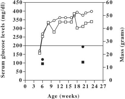
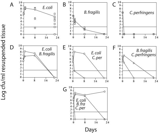
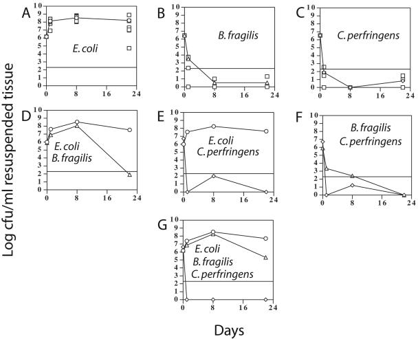
Human diabetics frequently suffer delayed wound healing, increased susceptibility to localized and systemic infections, and limb amputations as a consequence of the disease. Lower-limb infections in diabetic patients are most often polymicrobial, involving mixtures of aerobic, facultative anaerobic, and anaerobic bacteria. The purpose of this study is to determine if these organisms contribute to synergy in polymicrobial infections by using diabetic mice as an in vivo model. The model was the obese diabetic mouse strain BKS.Cg-m +/+ Lepr(db)/J, a model of human type 2 diabetes. Young (5- to 6-week-old) prediabetic mice and aged (23- to 24-week-old) diabetic mice were compared. The mice were injected subcutaneously with mixed cultures containing Escherichia coli, Bacteroides fragilis, and Clostridium perfringens. Progression of the infection (usually abscess formation) was monitored by examining mice for bacterial populations and numbers of white blood cells at 1, 8, and 22 days postinfection. Synergy in the mixed infections was defined as a statistically significant increase in the number of bacteria at the site of injection when coinfected with a second bacterium, compared to when the bacterium was inoculated alone. E. coli provided strong synergy to B. fragilis but not to C. perfringens. C. perfringens and B. fragilis provided moderate synergy to each other but only in young mice. B. fragilis was anergistic (antagonistic) to E. coli in coinfections in young mice at 22 days postinfection. When age-matched nondiabetic mice (C57BLKS/J) were used as controls, the diabetic mice exhibited 5 to 35 times the number of CFU as did the nondiabetic mice, indicating that diabetes was a significant factor in the severity of the polymicrobial infections.
Figures
Similar articles
-
Persistence of polymicrobial abscesses in the poorly controlled diabetic host.Diabetes. 1986 Apr;35(4):448-53. doi: 10.2337/diab.35.4.448. Diabetes. 1986. PMID: 3514325
-
The spectrum of Escherichia coli--Bacteroides fragilis pathogenic synergy in an intraabdominal infection model.Can J Microbiol. 1988 Mar;34(3):352-7. doi: 10.1139/m88-064. Can J Microbiol. 1988. PMID: 3046726
-
The treatment of irradiated mice with polymicrobial infection caused by Bacteroides fragilis and Escherichia coli.J Antimicrob Chemother. 1994 Feb;33(2):243-52. doi: 10.1093/jac/33.2.243. J Antimicrob Chemother. 1994. PMID: 8182005
-
Virulence factors of anaerobic bacteria.Johns Hopkins Med J. 1982 Jul;151(1):1-9. Johns Hopkins Med J. 1982. PMID: 7045489 Review. No abstract available.
-
Pathogenicity of the Bacteroides fragilis group.Ann Clin Lab Sci. 1989 Sep-Oct;19(5):360-76. Ann Clin Lab Sci. 1989. PMID: 2679351 Review.
Cited by
-
In vivo efficacy of humanized ceftaroline fosamil-avibactam exposures in a polymicrobial infection model.Antimicrob Agents Chemother. 2013 Nov;57(11):5674-8. doi: 10.1128/AAC.01162-13. Epub 2013 Sep 16. Antimicrob Agents Chemother. 2013. PMID: 24041891 Free PMC article.
-
An in vivo polymicrobial biofilm wound infection model to study interspecies interactions.PLoS One. 2011;6(11):e27317. doi: 10.1371/journal.pone.0027317. Epub 2011 Nov 4. PLoS One. 2011. PMID: 22076151 Free PMC article.
-
Polymicrobial Infections and Biofilms: Clinical Significance and Eradication Strategies.Antibiotics (Basel). 2022 Dec 1;11(12):1731. doi: 10.3390/antibiotics11121731. Antibiotics (Basel). 2022. PMID: 36551388 Free PMC article. Review.
-
The dynamic wound microbiome.BMC Med. 2020 Nov 24;18(1):358. doi: 10.1186/s12916-020-01820-6. BMC Med. 2020. PMID: 33228639 Free PMC article. Review.
-
Anaerobes in diabetic foot infections: pathophysiology, epidemiology, virulence, and management.Clin Microbiol Rev. 2024 Sep 12;37(3):e0014323. doi: 10.1128/cmr.00143-23. Epub 2024 May 31. Clin Microbiol Rev. 2024. PMID: 38819166 Free PMC article. Review.
References
-
- Apelqvist, J., and J. Larsson. 2000. What is the most effective way to reduce incidence of amputation in the diabetic foot? Diabetes-Metab. Res. Rev. 16:S75-S83. - PubMed
-
- Bessman, A. N., F. L. Sapico, M. Tabatabai, and J. Z. Montgomerie. 1986. Persistence of polymicrobial abscesses in the poorly controlled diabetic host. Diabetes 35:448-453. - PubMed
-
- Bjornson, H. S., R. Colley, R. H. Bower, V. P. Duty, J. T. Schwartz-Fulton, and J. E. Fischer. 1982. Association between microorganism growth at the catheter insertion site and colonization of the catheter in patients receiving total parenteral nutrition. Surgery 92:720-727. - PubMed
-
- Boquet, P. 2001. The cytotoxic necrotizing factor 1 (CNF1) from Escherichia coli. Toxicon 39:1673-1680. - PubMed
Publication types
MeSH terms
LinkOut - more resources
Full Text Sources
Medical
Molecular Biology Databases
Miscellaneous
