. 2005 Sep;73(9):6127-37.
doi: 10.1128/IAI.73.9.6127-6137.2005.

Anti-LcrV antibody inhibits delivery of Yops by Yersinia pestis KIM5 by directly promoting phagocytosis

Affiliations

Anti-LcrV antibody inhibits delivery of Yops by Yersinia pestis KIM5 by directly promoting phagocytosis

Clarissa Cowan et al. Infect Immun. 2005 Sep.

Abstract

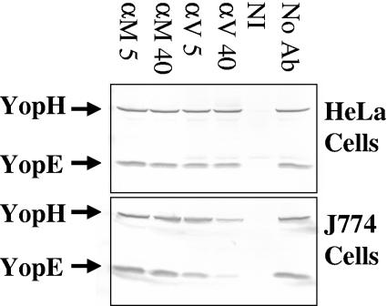
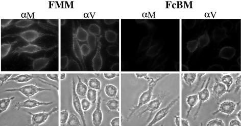
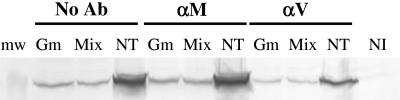
LcrV of Yersinia pestis is a major protective antigen proposed for inclusion in subunit plague vaccines. One way that anti-LcrV antibody is thought to protect is by inhibiting the delivery of toxins called Yops to host cells. The present study characterizes the relation between this inhibition and the phagocytosis of the bacteria. J774A.1 cells were infected with Y. pestis KIM5 in the presence of a protective polyclonal anti-LcrV antibody or a nonprotective polyclonal anti-YopM antibody, and delivery of YopH and YopE into the cytoplasm was assayed by immunoblotting. The ability to inhibit the delivery of these Yops depended upon having antibody bound to the cell surface; blocking conditions that prevented the binding of antibody to Fc receptors prevented the inhibition of Yop delivery. Anti-LcrV antibody also promoted phagocytosis of the yersiniae, whereas F(ab')(2) fragments did not. Further, anti-LcrV antibody could not inhibit the delivery of Yops into cells that were unable to phagocytose due to the presence of cytochalasin D. However, Yops were produced only by extracellular yersiniae. We hypothesize that anti-LcrV antibody does not directly inhibit Yop delivery but instead causes phagocytosis, with consequent inhibition of Yop protein production in the intracellular yersiniae. The prophagocytic effect of anti-LcrV antibody extended to mouse polymorphonuclear neutrophils (PMNs) in vitro, and PMNs were shown to be critical for protection: when PMNs in mice were ablated, the mice lost all ability to be protected by anti-LcrV antibody.


Figures

FIG. 1.
FIG. 1.
Anti-LcrV antibody partially inhibits the delivery of YopH and YopE to J774A.1 cells but has no effect on the delivery of these Yops to HeLa cells. HeLa and J774A.1 cells were infected at an MOI of 10 with 26/37°C-grown Y. pestis KIM5 in the presence of anti-YopM (αM) or anti-LcrV (αV) antibody and the mild HgG blocking agent. After 4 h, delivery of YopH and YopE to the cytosol was measured by immunoblotting. Lanes: αM and αV 5, 5 μg/ml antibody; αM and αV 40, 40 μg/ml antibody; NI, noninfected; No Ab, cells were infected in the absence of antibody.
FIG. 2.
FIG. 2.
Effects of two blocking treatments on the binding of anti-LcrV and anti-YopM antibody to J774A.1 cells. Binding of anti-YopM (αM) or anti-LcrV (αV) antibody to J774A.1 cells was determined for two blocking regimens. Then the cells were treated with Oregon Green 488-conjugated secondary antibody in PBS and visualized by fluorescence (top panels) or phase-contrast (bottom panels) microscopy. FMM, 10% FBS with 1% MS pretreatment for 30 min, and then 40 μg/ml αM or αV in the presence of 1% MS; FcBM, FcBlock (rat monoclonal antibody against a common epitope in the extracellular domains of FcγRII and FcγRIII) with 1% BSA pretreatment for 30 min, and then 40 μg/ml αM or αV in the presence of 1% MS.
FIG. 3.
FIG. 3.
Specific blocking of Fc receptors on J774A.1 cells abrogates the inhibition of YopH delivery by anti-LcrV antibody. Panel A: control test for efficacy of FcBlock in inhibiting phagocytosis mediated by IgG1. J774A.1 cells were treated with Fc Block or PBS for 1 h and then allowed to engulf opsonized fluorescent beads for 1 h. The image shows an overlay of the fluorescent image of the beads onto the phase-contrast image to reveal the cellular outlines. Panel B: J774A.1 cells were given the indicated blocking regimens and infected with Y. pestis KIM5 for 4 h at 37°C in the presence of 40 μg/ml anti-YopM (αM) or anti-LcrV (αV) antibody. Delivery of YopH into the J774A.1 cells was determined by immunoblotting of samples of the soluble cytoplasmic fraction. Blocking treatments: FcBM, RPMI 1640 plus FcBlock plus 1% BSA pretreatment for 1.25 h at 37°C and then infection with yersiniae suspended in RPMI 1640 containing αM or αV and 1% MS; FMM, RPMI 1640 plus 10% FBS plus 1% MS pretreatment for 1.25 h at 37°C and then infection with yersiniae suspended in RPMI 1640 containing αM or αV and 1% MS; None, pretreatment with RPMI 1640 at 37°C for 1.25 h followed by infection with yersiniae suspended in RPMI 1640 containing αM or αV.
FIG. 4.
FIG. 4.
Anti-LcrV antibody can delay cytotoxicity in infected HeLa cells if no blocking agent is present. HeLa cells given the indicated blocking regimens were infected with Y. pestis KIM5 at an MOI of 2 in RPMI 1640 containing 40 μg/ml anti-YopM antibody (αM), 40 μg/ml anti-LcrV antibody (αV), or no antibody (No Ab). At various times after infection, cytotoxicity due to delivery of Yops was assessed by phase-contrast microscopy. The images shown were taken at 3 h postinfection. For blocking with HgG, the HeLa cells were pretreated for 30 min at 37°C with RPMI 1640 containing 500 μg/ml HgG and then were infected with yersiniae in RPMI 1640 containing αM or αV and 50 μg/ml HgG; for experiments with no blocking agent, the cells were pretreated in RPMI 1640 and infected with yersiniae in RPMI 1640 containing anti-YopM or anti-LcrV antibody. Ab, antibody; NI, not infected.
FIG. 5.
FIG. 5.
Full-length anti-LcrV antibody, but not F(ab′)2 fragments, promotes the phagocytosis of Y. pestis KIM5 pVGFP in J774A.1 cells. J774A.1 cells were infected at an MOI of 3 with Y. pestis KIM5 pVGFP in the presence of 40 μg/ml anti-LcrV, 100 μg/ml anti-LcrV F(ab′)2, or no antibody. After 1 h, the cells were fixed, and extracellular yersiniae were stained with anti-Yersinia primary antibodies and Texas Red-conjugated secondary antibody. Intracellular bacteria would not be stained and were green due to the expression of GFP. Numbers of extracellular and intracellular yersiniae were counted in 20 cells in 20 fields. Panel A shows fluorescence micrographs from such assays to illustrate the discrimination of intracellular and extracellular yersiniae. αLcrV, anti-LcrV; αYopM, anti-YopM. Panel B shows the data obtained for F(ab′)2 fragments of anti-LcrV antibody [αV F(ab′)2], PBS (No Ab), and anti-LcrV antibody (αV). Filled bars: intracellular yersiniae; open bars: extracellular yersiniae. Error bars show ±1 standard deviation (SD) from the mean numbers per J774A.1 cell. The results from the treatment with F(ab′)2 fragments of anti-LcrV antibody differed significantly from those from the treatment with full-length anti-LcrV at a P value of 0.05 (*).
FIG. 5.
FIG. 5.
Full-length anti-LcrV antibody, but not F(ab′)2 fragments, promotes the phagocytosis of Y. pestis KIM5 pVGFP in J774A.1 cells. J774A.1 cells were infected at an MOI of 3 with Y. pestis KIM5 pVGFP in the presence of 40 μg/ml anti-LcrV, 100 μg/ml anti-LcrV F(ab′)2, or no antibody. After 1 h, the cells were fixed, and extracellular yersiniae were stained with anti-Yersinia primary antibodies and Texas Red-conjugated secondary antibody. Intracellular bacteria would not be stained and were green due to the expression of GFP. Numbers of extracellular and intracellular yersiniae were counted in 20 cells in 20 fields. Panel A shows fluorescence micrographs from such assays to illustrate the discrimination of intracellular and extracellular yersiniae. αLcrV, anti-LcrV; αYopM, anti-YopM. Panel B shows the data obtained for F(ab′)2 fragments of anti-LcrV antibody [αV F(ab′)2], PBS (No Ab), and anti-LcrV antibody (αV). Filled bars: intracellular yersiniae; open bars: extracellular yersiniae. Error bars show ±1 standard deviation (SD) from the mean numbers per J774A.1 cell. The results from the treatment with F(ab′)2 fragments of anti-LcrV antibody differed significantly from those from the treatment with full-length anti-LcrV at a P value of 0.05 (*).
FIG. 6.
FIG. 6.
YopH is not delivered into J774A.1 cells by intracellular Y. pestis. Three sets of three J774A.1 cultures were infected with Y. pestis KIM5 in the presence of 40 μg/ml anti-LcrV antibody (αV), anti-YopM antibody (αM), or no antibody (No Ab). After 30 min, one culture of each group received Gm, one received a mixture of antibiotics at high concentration (Mix), and one was not treated with any antibiotics (NT). The molecular weight (mw) of the prestained marker shown was 47,500; an extract from a noninfected J774A.1 culture (NI) also was included. After 1 h, the cells were washed and given fresh medium lacking any antibiotics but containing antibodies as described above, and incubation was continued for an additional 2.5 h. The cytoplasmic fraction of the J774A.1 cells was recovered and analyzed for YopH by immunoblotting.
FIG. 7.
FIG. 7.
Functional actin is required for anti-LcrV antibody to inhibit the delivery of YopH and YopE into J774A.1 cells. J774A.1 cells pretreated with different concentrations of cytochalasin D (Cyto D) were infected with Y. pestis KIM5 for 4 h in the presence of 40 μg/ml anti-LcrV antibody (αV), anti-YopM antibody (αM), or no antibody (No Ab), and delivery of YopH and YopE into the soluble cytoplasmic fraction was assayed by immunoblotting.
FIG. 8.
FIG. 8.
Anti-Yersinia antibody also reduces the delivery of YopH and YopE. J774A.1 cells in six-well cluster dishes were infected at an MOI of 10 with Y. pestis KIM5(pGFP) in the presence of 40 μg/ml anti-LcrV or anti-Yersinia antibody or of PBS. Delivery of YopH and YopE to the soluble J774A.1 cellular fraction was assayed by immunoblotting. Lanes: PBS, no antibody; αYersinia, anti-Yersinia antibody; α-V, anti-LcrV antibody.
FIG. 9.
FIG. 9.
PMNs are essential for early protection by anti-LcrV antibody. C57BL/6 mice were given the anti-Ly-6G monoclonal antibody 1A8 at −18 h and on days 1 and 2 postinfection to ablate PMNs. Groups of these mice and of control mice whose PMNs were not ablated were also treated with a protective 100-μg dose of anti-LcrV antibody at −18 h. Additional control C57BL/6 mice were given a mock treatment with PBS at this time. All mice were infected retroorbitally (intravenously) on day 0 with 3 × 10⁴ Y. pestis KIM5 organisms. Groups of three mice per treatment were analyzed for CFU counts in the liver and spleen on days 2 and 3 postinfection. Open bars, control mice given anti-LcrV antibody; filled bars, control mice given PBS; light-grey bars, PMN-depleted mice given anti-LcrV antibody; dark-grey bars, PMN-depleted mice given PBS. Error bars indicate ±1 SD from the mean. PMN-depleted mice given PBS died before the 72-h point (†). Viable numbers in PMN-depleted mice given anti-LcrV antibody differed significantly from those in control mice given anti-LcrV antibody at P values of <0.05 (*) and <0.01 (**).
FIG. 10.
FIG. 10.
Anti-LcrV antibody promotes phagocytosis by PMNs. Glycogen-elicited PMNs from C57BL/6 mice were infected in suspension at an MOI of 3 with Y. pestis KIM5 pVGFP in the presence of 40 μg/ml anti-LcrV antibody (αV) or PBS (No Ab). Phagocytosis was assayed after 1 h as described in the legend to Fig. 5. Numbers of extracellular and intracellular yersiniae were counted in 20 cells in 20 fields. Filled bars, intracellular yersiniae; open bars, extracellular yersiniae. Error bars show ±1 SD from the mean numbers per PMN. The data for αV treatment differed significantly from those for PBS at a P value of <0.0001.

Similar articles

Cited by

References

    1. Bergman, T., S. Håkansson, Å. Forsberg, L. Norlander, A. Macellaro, A. Bäckman, I. Bölin, and H. Wolf-Watz. 1991. Analysis of the V antigen lcrGVH-yopBD operon of Yersinia pseudotuberculosis: evidence for a regulatory role of LcrH and LcrV. J. Bacteriol. 173:1607-1616. - PMC - PubMed
    1. Brubaker, R. R. 2003. Interleukin-10 and inhibition of innate immunity to yersiniae: roles of Yops and LcrV (V antigen). Infect. Immun. 71:3673-3681. - PMC - PubMed
    1. Cormack, B. P., R. H. Valdivia, and S. Falkow. 1996. FACS-optimized mutants of the green fluorescent protein (GFP). Gene 173:33-38. - PubMed
    1. Cornelis, G. R. 2002. Yersinia type III secretion: send in the effectors. J. Cell Biol. 158:401-408. - PMC - PubMed
    1. Cowan, C., H. A. Jones, Y. H. Kaya, R. D. Perry, and S. C. Straley. 2000. Invasion of epithelial cells by Yersinia pestis: evidence for a Y. pestis-specific invasin. Infect. Immun. 68:4523-4530. - PMC - PubMed

Publication types

MeSH terms

Substances