

Vitrectomy and gas tamponade without internal limiting membrane peeling for myopic foveoschisis
- PMID: 16113377
- PMCID: PMC1772841
- DOI: 10.1136/bjo.2005.069427
Vitrectomy and gas tamponade without internal limiting membrane peeling for myopic foveoschisis
Abstract
Aim: To evaluate the clinical and anatomical outcomes of pars plana vitrectomy and gas tamponade without internal limiting membrane (ILM) peeling in symptomatic patients caused by myopic foveoschisis.
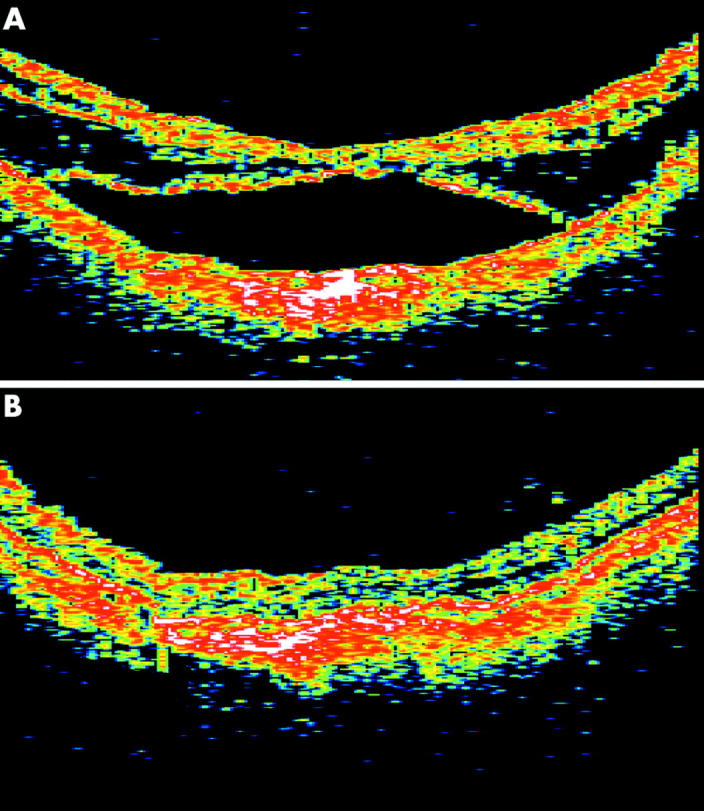
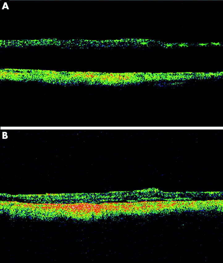
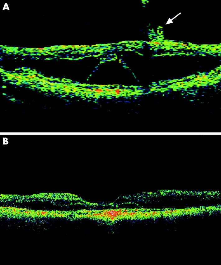
Methods: Nine eyes in eight highly myopic patients who had myopic foveoschisis with foveal detachment underwent vitrectomy without ILM peeling followed by gas tamponade. Main outcome measures include change in best corrected visual acuity (BCVA) and changes in height of the foveal detachment and resolution of the myopic foveoschisis measured by optical coherence tomography (OCT).
Results: After surgery, BCVA improved in eight eyes with the median BCVA improved from 20/80 to 20/50 (p=0.012). The mean line of visual improvement was 3.6 lines. OCT showed complete resolution of myopic foveoschisis with complete foveal reattachment in seven (77.8%) eyes with partial resolution in two (22.2%) eyes. The mean height of foveal detachment decreased from 505 mum preoperatively to 21 mum postoperatively (p<0.001).
Conclusions: Vitrectomy without ILM peeling followed by gas tamponade appeared to result in favourable visual and anatomical outcomes for treating myopic foveoschisis in highly myopic eyes. The results are comparable with studies in which ILM removal was performed. Further controlled study will be useful to determine the role of ILM peeling in these patients.
Figures
References
-
- Takano M , Kishi S. Foveal retinoschisis and retinal detachment in severely myopic eyes with posterior staphyloma. Am J Ophthalmol 1999;128:472–6. - PubMed
-
- Ikuno Y , Sayanagi K, Ohji M, et al. Vitrectomy and internal limiting membrane peeling for myopic foveoschisis. Am J Ophthalmol 2004;137:719–24. - PubMed
-
- Baba T , Ohno-Matsui K, Futagami S, et al. Prevalence and characteristics of foveal retinal detachment without macular hole in high myopia. Am J Ophthalmol 2003;135:338–42. - PubMed
-
- Panozzo G , Mercanti A. Optical coherence tomography findings in myopic traction maculopathy. Arch Ophthalmol 2004;122:1455–60. - PubMed
-
- Akiba J , Konno S, Sato E, et al. Retinal detachment and retinoschisis detected by optical coherence tomography in a myopic eye with a macular hole. Ophthalmic Surg Lasers 2000;31:240–2. - PubMed
MeSH terms
Substances
LinkOut - more resources
Full Text Sources
Miscellaneous