. 2005 Sep;7(9):901-8.
doi: 10.1038/ncb1293. Epub 2005 Aug 21.

Phospho-caveolin-1 mediates integrin-regulated membrane domain internalization

Affiliations

Phospho-caveolin-1 mediates integrin-regulated membrane domain internalization

Miguel A del Pozo et al. Nat Cell Biol. 2005 Sep.

Abstract

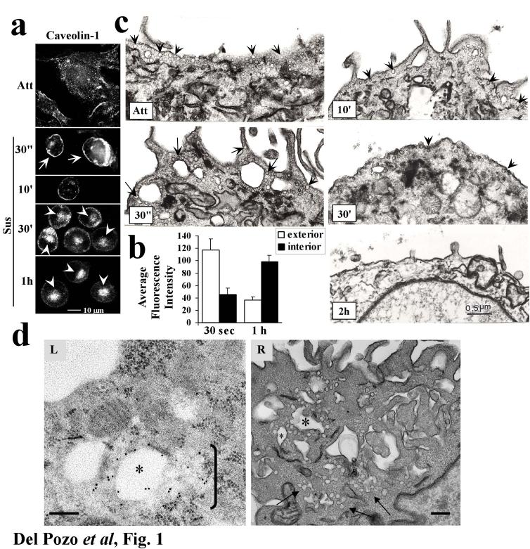
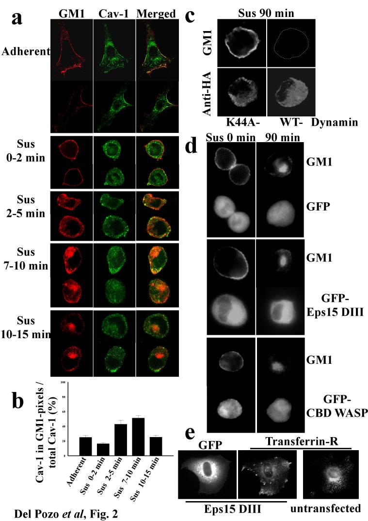
Growth of normal cells is anchorage dependent because signalling through multiple pathways including Erk, phosphatidylinositol-3-OH kinase (PI(3)K) and Rac requires integrin-mediated cell adhesion. Components of these pathways localize to low-density, cholesterol-rich domains in the plasma membrane named 'lipid rafts' or 'cholesterol-enriched membrane microdomains' (CEMM). We previously reported that integrin-mediated adhesion regulates CEMM transport such that cell detachment from the extracellular matrix triggers CEMM internalization and clearance from the plasma membrane. We now report that this internalization is mediated by dynamin-2 and caveolin-1. Internalization requires phosphorylation of caveolin-1 on Tyr 14. A shift in localization of phospho-caveolin-1 from focal adhesions to caveolae induces CEMM internalization upon cell detachment, which mediates inhibition of Erk, PI(3)K and Rac. These data define a novel molecular mechanism for growth and tumour suppression by caveolin-1.


Figures

Figure 1
Figure 1
Caveolin-1 internalisation. (a) Adherent 3T3 cells or cells suspended for the indicated times were fixed, permeabilized and stained for caveolin-1. Cell surface (arrows) vs. intracellular (arrowheads) staining is indicated. (b) Internalisation of caveolin in images from (a) was quantified as described in Methods. Caveolin fluorescence within 3.5 microns of the cell surface is considered exterior; fluorescence internal to this zone, further from the plasma membrane, is considered interior. Values are means ± S.E.M. from 10 cells in 3 independent experiments. Differences between 30s and 1h are statistically significant (p<2.5×10⁻⁴ for the interior region and p<7×10⁻³ for the exterior). (c) Attached cells or cells suspended for the indicated times were fixed and processed for electron microscopy to detect caveolae in the plasma membrane. Caveolae are indicated by arrows. Images are representative of areas within cells where caveolae are concentrated. (d) Cells suspended for 2 min. were fixed and processed for either (L) immunogold labelling of caveolin-1 or (R) electron microscopy. (L) A region of caveolae internalisation showing a vacuole surrounded by multiple α-Cav-1 IgG gold particles. To the right of this vacuole (bracket) is a cluster of caveolae emanating from the vacuole. (R) A similar region of a cell processed for regular TEM microscopy. This image shows several caveolae-rich vacuoles (asterisks) and associated clusters of caveolae and caveolae-derived vesicles (arrows). Bar=0.2 μm. (n=3).
Figure 2
Figure 2
Pathway of integrin-regulated GM1 internalisation. (a) Serum deprived NIH-3T3 fibroblasts were surface labelled on ice with CTxB-Alexa 568, detached and held in suspension (Sus) for the indicated times, or plated on FN for 2hours (Adherent). Cells were then fixed and stained for caveolin-1 in green. Cells shown are observed at the equatorial plane and representative of up to 50 cells at each time point. (b) To quantify co-localization between GM-1 and caveolin, pixels that were positive for GM1 stain were identified and the fraction of the caveolin-1 stain within these zones calculated. Values are means ± S.E.M. from 3 independent experiments. (c) wt MEFs expressing HA-tagged dominant negative (K44A) or wt dynamin-2 were held in suspension for 90 min, surface labelled with Alexa 488-CTxB, and then stained with anti-HA antibody. (d) wt MEFs expressing GFP alone, GFP-tagged Eps15DIII, or the Cdc42-binding domain (CBD) of WASP were surface labelled with Alexa 568-CTB, then detached and held in suspension for the indicated times. (e) Control and GFP-tagged Eps15DIII expressing wt MEFs adherent on FN were stained for the transferrin receptor. Cells shown are representative of three independent experiments.
Figure 3
Figure 3
Requirement for Caveolin-1. (a) Lysates from M21L melanoma or M21L-cav cells were analysed by Western blotting with anti-caveolin polyclonal Ab. (b) Cells detached for 2h (top panels) were stained live with FITC-CTxB to label surface GM1. Middle panels: cells expressing GFP-V12Rac. Bottom panels: Cells stained with rhodamine-phalloidin. Arrows indicate protrusions and ruffle-like structures (c) GFPV12Rac localization to the membrane in (b) was assessed by measuring pixel intensity starting at the cell edge. Values are means ± SEM. (d) Confocal z-section series of actin-stained cells. (e) Serum-starved caveolin-1-/- MEFs (KO) or caveolin-1+/+ MEFs (WT), adherent or suspended for 2h, were stimulated with 10% serum for 10 min, and PAK kinase activity assayed. (f) Caveolin-1-/- MEFs (KO) or caveolin-1+/+ MEFs (WT) were kept adherent or suspended for 8h in the presence of 10% serum. Cell lysates were analysed by Western blotting with phospho-specific antibodies to Erk1/2, Akt, and FAK, or with phospho-independent antibodies to the same proteins for normalization. Values in (e) and (f) are means ± S.D. of the specific enzymatic activity (kinase activity or amount of phospho-protein normalized to total protein) in non-adherent cells normalized to that of adherent cells (n=4).
Figure 4
Figure 4
Phospho-caveolin-1 mediates integrin-dependent membrane domain internalisation. (a) Adherent NIH-3T3 cells were doubly stained for phospho-caveolin-1 plus either vinculin or total caveolin-1. wt MEFs (b) or caveolin-1-/- MEFs (c) in suspension were stained for total caveolin-1 plus pY14cav-1. (d) NIH-3T3 cells were kept adherent or placed in suspension for the indicated times. Cell lysates were analysed by Western blotting with anti-caveolin-1 or anti-phosphocaveolin-1 (n=10). (e) Western blotting for caveolin-1 and flag in caveolin-1 +/+ MEFs (WT), caveolin-1 -/- MEFs (KO), and KO MEFs transiently transfected with flag-tagged WT or Y14F caveolin-1. (f). Caveolin-null MEFs transiently transfected with WT or Y14F caveolin-1 were detached for 2h and stained live with FITC-CTxB to label surface GM1, then fixed and stained with anti-flag mAb to detect caveolin expression. Arrows denote transfected cells. Images are single confocal sections (n=4). (g) Quantitation of surface GM1. The level of surface GM1 was quantified by measuring total staining intensity of suspended, surface CTxB-labelled untransfected caveolin-null cells and cells expressing WT or Y14F caveolin from (f). Values are means ± S.E.M. from 10 cells in 4 independent experiments. (h) NIH-3T3 cells, untreated COS-7 cells, and COS-7 cells incubated with 1μM sodium pervanadate for 1 h were lysed, and analysed by western blotting with antibodies against caveolin-1, pY14cav-1, and β1 integrin. (i) Adherent and suspended cells treated as in (h) were surface labelled with CTxB-Alexa 568. Asterisks denote cells not readily visible due to low surface labelling. Results are representative of three independent experiments.

References

    1. Schwartz MA. Integrins, oncogenes, and anchorage independence. J. Cell Biol. 1997;139:575–578. - PMC - PubMed
    1. Edidin M. The state of lipid rafts: from model membranes to cells. Annu Rev Biophys Biomol Struct. 2003;32:257–83. - PubMed
    1. Lagerholm BC, Weinreb GE, Jacobson K, Thompson NL. Detecting Microdomains in Intact Cell Membranes. Annu Rev Phys Chem. 2005;56:309–336. - PubMed
    1. Grande A, Echarri A, del Pozo MA. Integrin regulation of membrane domain trafficking and Rac targeting. Biochemical Society Transactions. 2005;33:609–613. - PubMed
    1. del Pozo MA, et al. Integrins regulate Rac targeting by internalization of membrane domains. Science. 2004;303:839–42. - PubMed

Publication types

MeSH terms