. 1992 Jan 31;571(1):50-63.
doi: 10.1016/0006-8993(92)90508-7.

Pontine regulation of REM sleep components in cats: integrity of the pedunculopontine tegmentum (PPT) is important for phasic events but unnecessary for atonia during REM sleep

Affiliations

Pontine regulation of REM sleep components in cats: integrity of the pedunculopontine tegmentum (PPT) is important for phasic events but unnecessary for atonia during REM sleep

M N Shouse et al. Brain Res. .

Abstract

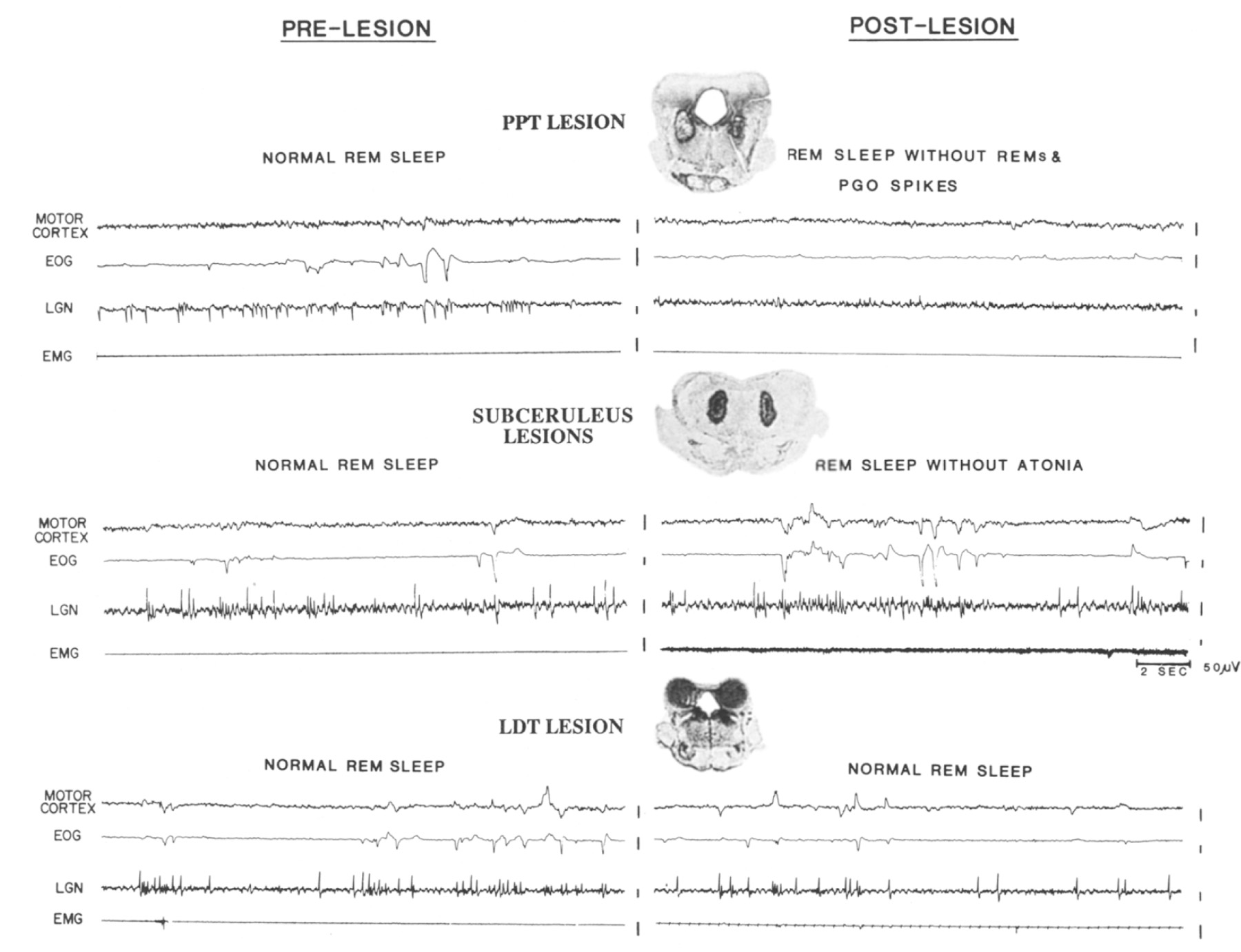
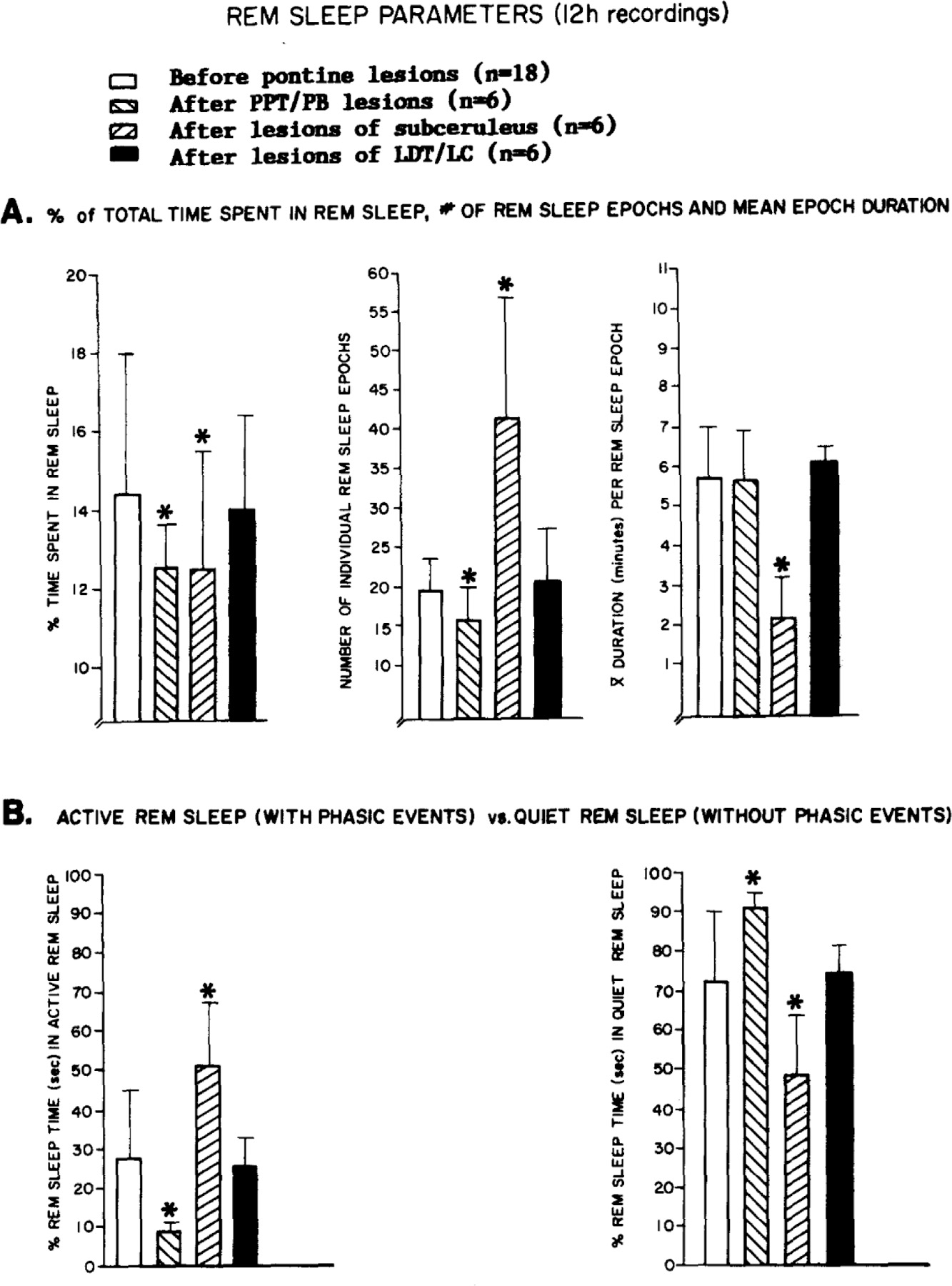
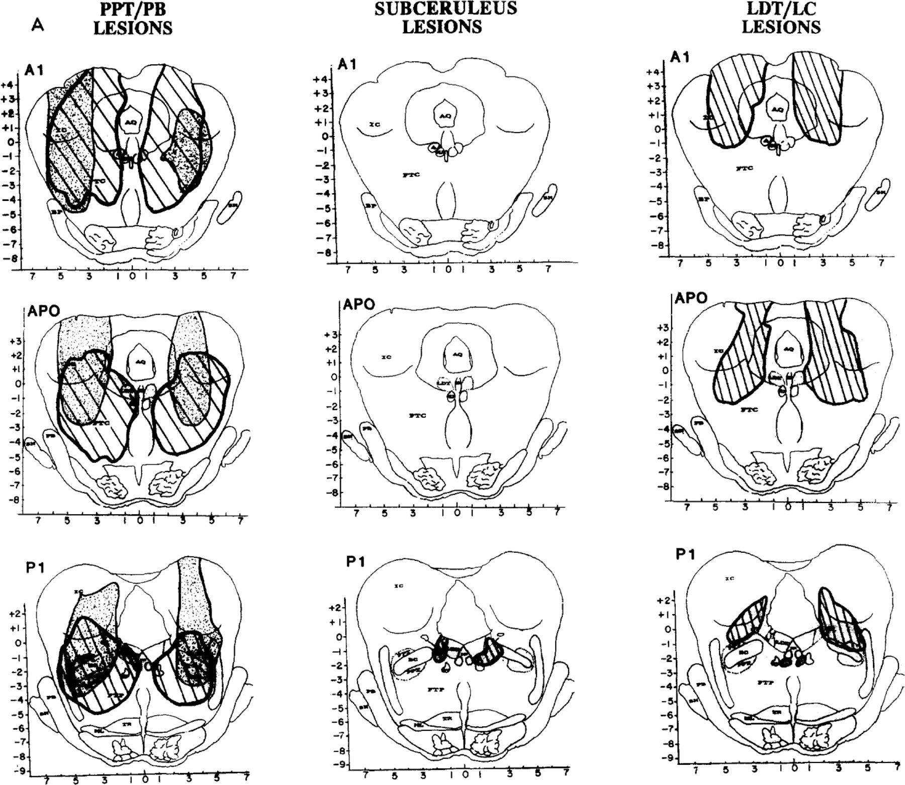
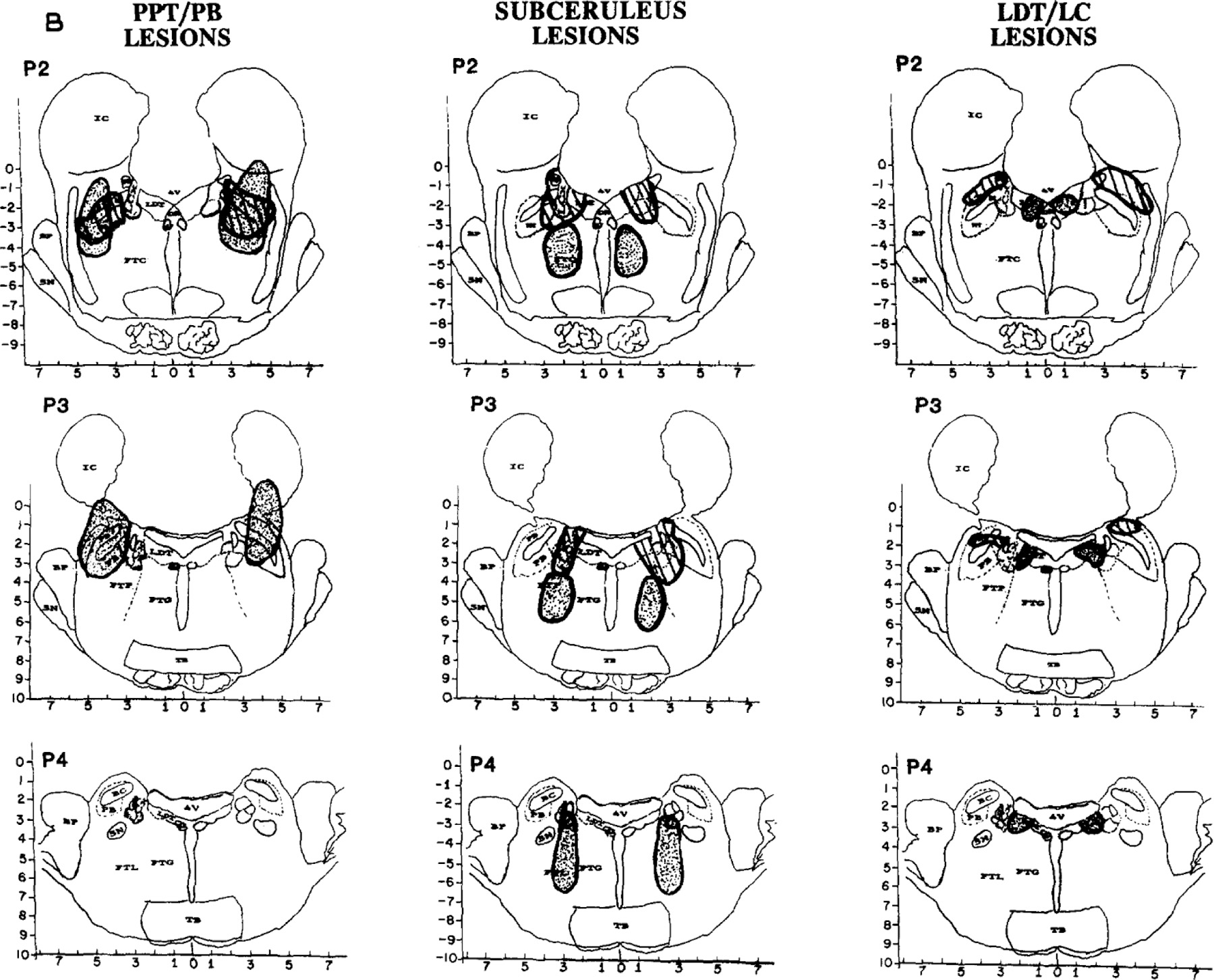
Transection, lesion and unit recording studies have localized rapid eye movement (REM) sleep mechanisms to the pons. Recent work has emphasized the role of pontine cholinergic cells, especially those of the pedunculopontine tegmentum (PPT). The present study differentiated REM sleep deficits associated with lesions of the PPT from other pontine regions implicated in REM sleep generation, including those with predominantly cholinergic vs non-cholinergic cells. Twelve hour polygraphic recordings were obtained in 18 cats before and 1-2 weeks after bilateral electrolytic or radio frequency lesions of either: (1) PPT, which contains the dorsolateral pontine cholinergic cell column; (2) laterodorsal tegmental nucleus (LDT), which contains the dorsomedial pontine cholinergic cell column; (3) locus ceruleus (LC), which contains mostly noradrenergic cells; or (4) subceruleus (LC alpha, peri-LC alpha and the lateral tegmental field), which also contains predominantly noncholinergic cells. There were three main findings: (i) Only lesions of PPT and subceruleus significantly affected REM sleep time. These lesions produced comparable reductions in REM sleep time but influenced REM sleep components quite differently: (ii) PPT lesions, estimated to damage 90 +/- 4% of cholinergic cells, reduced the number of REM sleep entrances and phasic events, including ponto-geniculooccipital (PGO) spikes and rapid eye movements (REMs), but did not prevent complete atonia during REM sleep: (iii) Subceruleus lesions eliminated atonia during REM sleep. Mobility appeared to arouse the cat prematurely from REM sleep and may explain the brief duration of REM sleep epochs seen exclusively in this group. Despite the reduced amount of REM sleep, the total number of PGO spikes and REM sleep entrances increased over baseline values. Collectively, the results distinguish pontine loci regulating phasic events vs atonia. PPT lesions reduced phasic events, whereas subceruleus lesions created REM sleep without atonia. Severe REM sleep deficits after large pontine lesions, including PPT and subceruleus, might be explained by simultaneous production of both REM sleep syndromes. However, extensive loss of ACh neurons in the PPT does not disrupt REM sleep atonia.


Figures

Fig. 1.
Fig. 1.
Twenty-second polygraphic tracings of REM sleep before and after lesions, together with a coronal section through the center of the pontine lesion in 1 cat per group. Each tracing displays a motor cortex electroencephalogram (EEG), an electrooculogram (EOG), an EEG from lateral geniculate nucleus (LGN) to register pontogeniculooccipital (PGO) spikes and an electromyogram (EMG). Motor cortex EEG desynchronization was intact regardless of lesion site. Top: Group 1 PPTIPB lesions, illustrated by a radio frequency lesion of the pedunculopontine tegmentum (PPT), diminshed clustered phasic events (rapid-eye-movements or REMs and PGO spikes) during REM sleep. Middle: Group 2 subceruleus lesions, illustrated by an electrolytic lesion of pen LC-alpha and the underlying reticular core, mostly the lateral tegmental field (FTL), abolished atonia during REM sleep and also seemed to increase phasic events (REMs and PGO spikes). Bottom: Group 3 LDT and/or LC lesions, illustrated by a lesion encompassing nearly all of the laterodorsal tegmental nucleus (LDT), did not affect REM sleep.
Fig. 2.
Fig. 2.
Means and standard deviations for various parameters of REM sleep before and after lesions in 3 groups of cats. Two-way analysis of variance compared each variable as a function of lesion group (PPT/PB, subceruleus or LDT/LC) with repeated measures over time (pre-vs post-lesion). A: percent of the 12 h recording time spent in REM sleep (group: F = NS; time: F = 36.3, P < 0.01; group x time: F 12.8, P < 0.01), number of REM sleep episodes (group: F = 7.3, P < 0.01; time: F =8.1, P < 0.01, Group x time: F = 17.3, P < 0.05) and mean REM sleep epoch duration (group: F = 8.9, P < 0.01; time: F = 10.5, P < 0.01; group x time: F = 8,0, P < 0.05). 13: percentages of active vs quiet REM sleep. Active REM sleep is defined by clustered phasic events, whereas quiet REM sleep does not contain clustered REMs and PGOs. Each second of REM sleep was classified as active or quiet. F tests are identical, as active and quiet REM percentages cumulate to 100% (group: F = 3.95, P < 0.05; time: F = NS; group x time: F = 8.0, P < 0.05). Post hoc t-tests indicated no difference between groups in pre-lesion measures, which are combined. There were also no differences between pre- and post-lesion REM sleep measures in the LDT/LC group. Significant dependent or independent t-tests for PPT/PB or subceruleus lesion groups are indicated by asterisks.
Fig. 3.
Fig. 3.
B. Reconstructions of lesions in 2 cats (one dotted and the other striped) in each of the 3 lesion groups, PPT/PB (left), subceruleus (middle) and LDT/LC (right). Coronal section and abbreviations are adapted from the Berman atlas and approximate 1 mm intervals from Al throught P4. Left panel: Group 1 PPT/PB. Lesions were larger than in the other 2 groups and involved most of the lateral pontine cholinergic cell columns, including PFT and PB, often encompassed the central tegmental field (FTC) and sometimes the parvocellular tegmental field (FTP) as well. Tissue lost posterior to P2 is illustrated in 1 cat (dotted lesion); damage to the peribrachial (PB) cholinergic columns is evident at P3. Middle panel: Group 2 subceruleus. Lesions were smaller as well as caudal and ventromedial to PPT/PB lesions. Peri-LC alpha was damaged in all 6 cats. Some lesions encompassed locus ceruleus complex (LCx; striped lesion), and others damaged peri-LC alpha together with the underlying reticular formation (dotted), mostly the lateral tegemental field (FTL) and some of the gigantocellular tegmental field (FTG). Right panel: Group 3 LDT/LC. One cat in this group had a large lateral lesion involving most of FTC and some of the LC (striped). The other lesions in the group were more medial (dotted), encompassing all or parts of the lateral dorsal tegmenturn (LDT) and/or locus ceruleus (LC).
Fig. 3.
Fig. 3.
B. Reconstructions of lesions in 2 cats (one dotted and the other striped) in each of the 3 lesion groups, PPT/PB (left), subceruleus (middle) and LDT/LC (right). Coronal section and abbreviations are adapted from the Berman atlas and approximate 1 mm intervals from Al throught P4. Left panel: Group 1 PPT/PB. Lesions were larger than in the other 2 groups and involved most of the lateral pontine cholinergic cell columns, including PFT and PB, often encompassed the central tegmental field (FTC) and sometimes the parvocellular tegmental field (FTP) as well. Tissue lost posterior to P2 is illustrated in 1 cat (dotted lesion); damage to the peribrachial (PB) cholinergic columns is evident at P3. Middle panel: Group 2 subceruleus. Lesions were smaller as well as caudal and ventromedial to PPT/PB lesions. Peri-LC alpha was damaged in all 6 cats. Some lesions encompassed locus ceruleus complex (LCx; striped lesion), and others damaged peri-LC alpha together with the underlying reticular formation (dotted), mostly the lateral tegemental field (FTL) and some of the gigantocellular tegmental field (FTG). Right panel: Group 3 LDT/LC. One cat in this group had a large lateral lesion involving most of FTC and some of the LC (striped). The other lesions in the group were more medial (dotted), encompassing all or parts of the lateral dorsal tegmenturn (LDT) and/or locus ceruleus (LC).

References

    1. Aserinsky E and Kleitman N, Regularly occurring periods of eye motility, and concomitant phenomena during sleep, Science, 118 (1953) 273–274. - PubMed
    1. Baghdoyan HA, Monaco AR, Rodrigo-Angulo ML, Assens F, McCarley RW and Hobson JA, Microinjection of neostigmine into the pontine reticular formation of cats enhances desynchronized sleep signs, J. Pharmacol. Exp. Ther, 321 (1984) 173–180. - PubMed
    1. Baghdoyan HA, Rodrigo-Angulo ML, McCarley RW and Hobson JA, Site-specific enhancement and suppression of desynchronized sleep signs following cholinergic stimulation of three brainstem sites, Brain Research, 306 (1984) 39–52. - PubMed
    1. Berman AL, The Brain Stem of the Cat. A Cytoarchitectonic Atlas with Stereotaxic Coordinates University of Wisconsin Press, Madison, 1982.
    1. Carli G and Zanchetti A, A study of pontine lesions suppressing deep sleep in the cat, Arch. Ital. Biol, 103 (1965) 751–788. - PubMed

Publication types

Substances

LinkOut - more resources