. 2005 Oct 21;280(42):35647-57.
doi: 10.1074/jbc.M507924200. Epub 2005 Aug 22.

Retinoid absorption and storage is impaired in mice lacking lecithin:retinol acyltransferase (LRAT)

Affiliations

Retinoid absorption and storage is impaired in mice lacking lecithin:retinol acyltransferase (LRAT)

Sheila M O'Byrne et al. J Biol Chem. .

Abstract

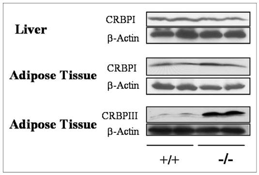
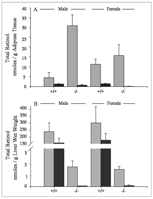
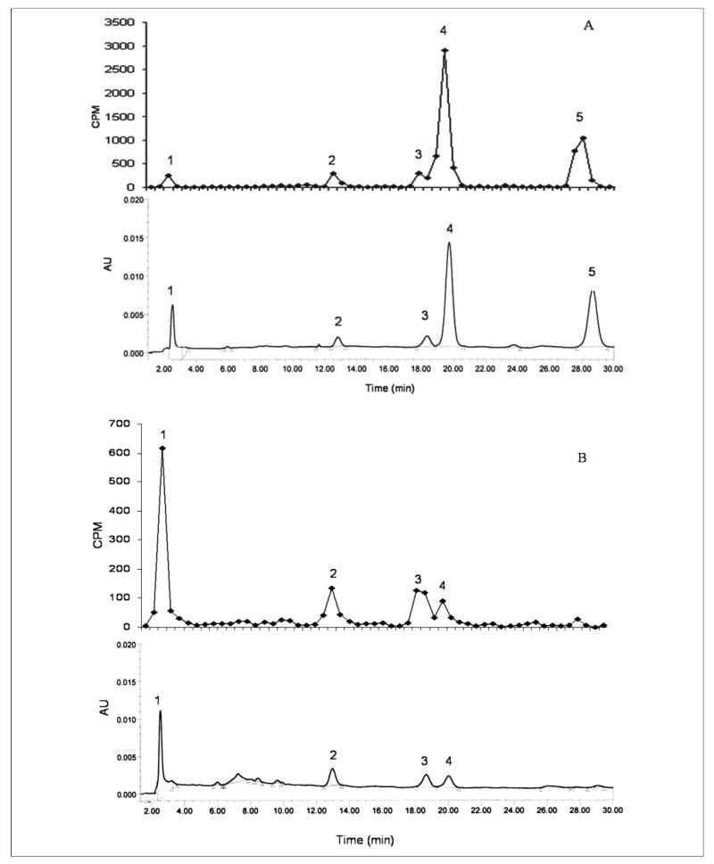
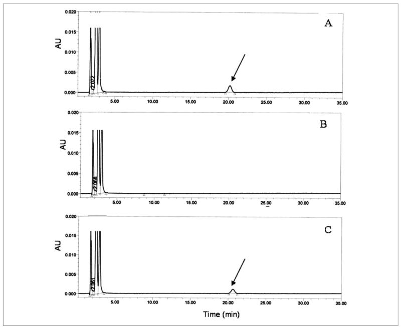
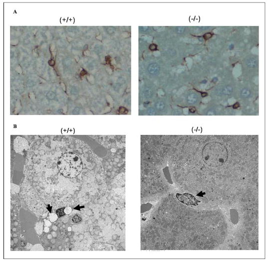
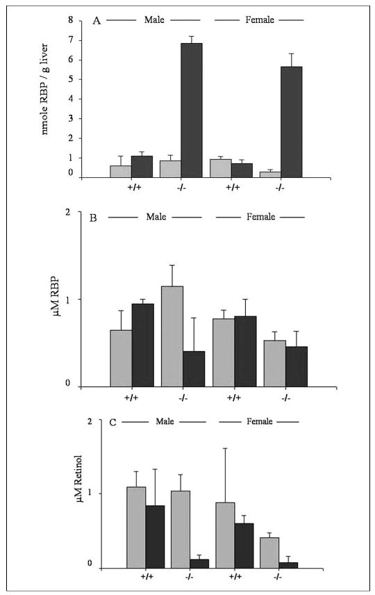
Lecithin:retinol acyltransferase (LRAT) is believed to be the predominant if not the sole enzyme in the body responsible for the physiologic esterification of retinol. We have studied Lrat-deficient (Lrat-/-) mice to gain a better understanding of how these mice take up and store dietary retinoids and to determine whether other enzymes may be responsible for retinol esterification in the body. Although the Lrat-/- mice possess only trace amounts of retinyl esters in liver, lung, and kidney, they possess elevated (by 2-3-fold) concentrations of retinyl esters in adipose tissue compared with wild type mice. These adipose retinyl ester depots are mobilized in times of dietary retinoid insufficiency. We further observed an up-regulation (3-4-fold) in the level of cytosolic retinol-binding protein type III (CRBPIII) in adipose tissue of Lrat-/- mice. Examination by electron microscopy reveals a striking total absence of large lipid-containing droplets that normally store hepatic retinoid within the hepatic stellate cells of Lrat-/- mice. Despite the absence of significant retinyl ester stores and stellate cell lipid droplets, the livers of Lrat-/- mice upon histologic analysis appear normal and show no histological signs of liver fibrosis. Lrat-/- mice absorb dietary retinol primarily as free retinol in chylomicrons; however, retinyl esters are also present within the chylomicron fraction obtained from Lrat-/- mice. The fatty acyl composition of these (chylomicron) retinyl esters suggests that they are synthesized via an acyl-CoA-dependent process suggesting the existence of a physiologically significant acyl-CoA:retinol acyltransferase.


Figures

FIGURE 1
FIGURE 1. CRBPIII but not CRBPI levels are elevated in adipose tissue from 3-month-old Lrat−/− mice.
Western blots for CRBPI in the liver and epididymal adipose tissue and for CRBPIII in epididymal adipose tissue for two male wild type (+/+) and two male Lrat−/− (−/−) mice are shown.
FIGURE 2
FIGURE 2. Effects of a totally retinoid-deficient diet on adipose tissue and liver levels of total retinol (retinol + retinyl ester) for wild type (+/+) and Lrat−/− (−/−) mice
Panel A shows the levels of total retinol (retinol + retinyl esters) in perigonadal adipose tissue obtained from male and female wild type and Lrat−/− mice. The groups receiving the control diet are indicated by light gray bars and the groups receiving the retinoid-deficient diet are indicated by dark gray bars. Panel B shows the levels of total retinol (retinol + retinyl esters) in the livers of male and female wild type and Lrat−/− mice receiving a control diet (light gray bars) or a retinoid-deficient diet (dark gray bars). All mice were maintained on a chow diet through the first 3 months of age, and at this point they were then randomly assigned to groups and maintained for 4 additional weeks on either the same chow diet or a totally retinoid-deficient purified diet.
FIGURE 3
FIGURE 3. Reverse phase HPLC profiles showing the distribution of retinol and retinyl esters and 3H counts/min present in chylomicrons obtained from wild type (panel A) and Lrat−/− (panel B) mice following administration of a gavage dose of retinol (6 μg containing 2 × 10⁶ 3H counts/min) in peanut oil
Panels A and B, the upper profiles show the distribution of [3H]retinoids and the lower profiles the UV absorbance of the retinoids. Note that for panels A and B the lower profiles are scaled to reflect at full scale the same absorbance units (AU). The extracted retinoids were separated on a 5-μm 4.6 × 250-mm Ultrasphere C18 column preceded by a C18 guard column, using 70% acetonitrile, 15% ethanol, 15% methylenechloride as the running solvent flowing at 1.8 ml/min. The numbers above the HPLC peaks indicate the following: 1, retinol; 2, retinyl linoleate; 3, retinyl oleate; 4, retinyl palmitate; and 5, retinyl stearate.
FIGURE 4
FIGURE 4. CRBPIII- but not CRBPI-bound retinol enables human DGAT1 catalysis of acyl-CoA-dependent esterification of retinol
Microsomes isolated from HEK293 cells transfected with a cDNA encoding human DGAT1 were incubated either in the presence of 16 μm free all-trans-retinol (panel A) or 16 μm all-trans-retinol bound to CRBPI (panel B) or 16 μm all-trans-retinol bound to CRBPIII (panel C) for 10 min. The HPLC peak identified with the arrow is that of all-trans-retinyl palmitate formed upon DGAT1 action.
FIGURE 5
FIGURE 5. The hepatic stellate cells of Lrat−/− mice lack lipid droplets that are a morphologic hallmark of these cells
Panel A shows the immunohistochemical staining pattern for desmin, a marker for hepatic stellate cells, in liver sections from 3-month-old male wild type (+/+) and Lrat−/− (−/−) mice. Panel B shows electron micrographs of liver sections prepared from 3-month-old male wild type and Lrat−/− mice. The electron micrographs show the presence of characteristic retinyl ester-containing lipid droplets in hepatic stellate cells of wild type mice (−/−) and their absence in livers from Lrat−/− mice (−/−). The arrows indicate the presence (+/+) or absence (−/−) of lipid droplets in hepatic stellate cells. The large adjoining cells are hepatocytes.
FIGURE 6
FIGURE 6. RBP levels are elevated in the livers of Lrat−/− mice but not wild type mice fed a totally retinoid-deficient diet for 1 month
Panel A shows RBP concentrations in livers from male and female wild type (+/+) and Lrat−/− (−/−) mice receiving either a nutritionally complete control diet (light gray bars) or a totally retinoid-deficient diet (dark gray bars). Panel B shows RBP concentrations in serum from male and female wild type (+/+) and Lrat−/− (−/−) mice receiving either a nutritionally complete control diet (light gray bars) or a totally retinoid-deficient diet (dark gray bars). Panel C shows serum retinol concentrations for male and female wild type (+/+) and Lrat−/− (−/−) mice receiving either a nutritionally complete control diet (light gray bars) or a totally retinoid-deficient diet (dark gray bars).

References

    1. Moore, T. (1957) Vitamin A, Elsevier Publishing Co., Amsterdam
    1. Wald G. Nature. 1968;219:800–807. - PubMed
    1. McBee JK, Palczweski K, Baehr W, Pepperberg DR. Prog Retin Eye Res. 2001;20:469–529. - PubMed
    1. Gudas, L. J., Sporn, M. B., and Roberts, A. B. (1994) in The Retinoids, Biology, Chemistry and Medicine (Sporn, M. B., Roberts, A. B., and Goodman, D. S., eds) 2nd Ed., pp. 443–520, Raven Press, Ltd., New York
    1. Balmer JE, Blomhoff R. J Lipid Res. 2002;43:1773–1808. - PubMed

Publication types

MeSH terms