. 2005 Aug 30;102(35):12519-24.
doi: 10.1073/pnas.0505641102. Epub 2005 Aug 22.

Atypical PKCiota contributes to poor prognosis through loss of apical-basal polarity and cyclin E overexpression in ovarian cancer

Affiliations

Atypical PKCiota contributes to poor prognosis through loss of apical-basal polarity and cyclin E overexpression in ovarian cancer

Astrid M Eder et al. Proc Natl Acad Sci U S A. .

Abstract

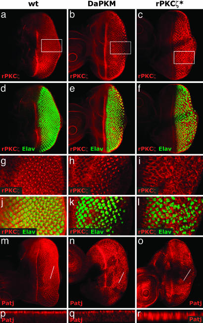
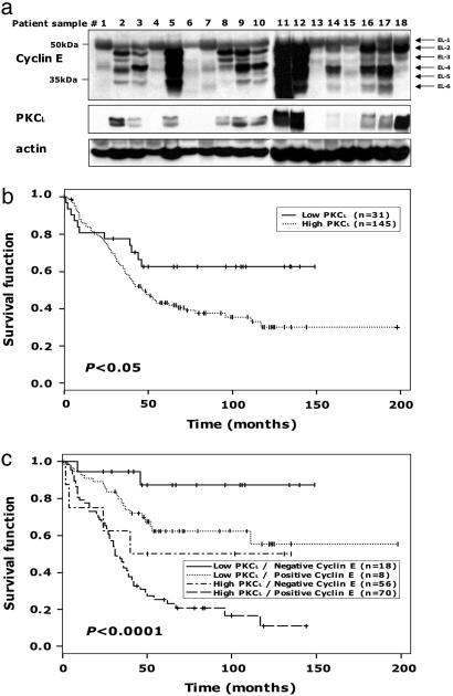
We show that atypical PKCiota, which plays a critical role in the establishment and maintenance of epithelial cell polarity, is genomically amplified and overexpressed in serous epithelial ovarian cancers. Furthermore, PKCiota protein is markedly increased or mislocalized in all serous ovarian cancers. An increased PKCiota DNA copy number is associated with decreased progression-free survival in serous epithelial ovarian cancers. In a Drosophila in vivo epithelial tissue model, overexpression of persistently active atypical PKC results in defects in apical-basal polarity, increased Cyclin E protein expression, and increased proliferation. Similar to the Drosophila model, increased PKCiota proteins levels are associated with increased Cyclin E protein expression and proliferation in ovarian cancers. In nonserous ovarian cancers, increased PKCiota protein levels, particularly in the presence of Cyclin E, are associated with markedly decreased overall survival. These results implicate PKCiota as a potential oncogene in ovarian cancer regulating epithelial cell polarity and proliferation and suggest that PKCiota is a novel target for therapy.


Figures

Fig. 1.
Fig. 1.
Amplification of the PKCι gene and increased PKCι RNA expression in ovarian cancer. (a) Array comparative genomic hybridization analysis of PKCι DNA copy number in 235 Grade 3 and Stage III or IV serous epithelial ovarian cancer samples (log2 ratio of cancer patient DNA to normal DNA). (b) Increase in PKCι DNA copy number is associated with a decreased progression-free survival period. For patients where followup information was available, progression-free survival in patients with high PKCι DNA copy number (n = 26) was significantly worse (P = 0.0006) than in patients with low PKCι copy number (n = 46) (cutoff at 0.37 log2). Vertical lines indicate censored patients, i.e., patients for whom no further followup information was available after the indicated time points. (c) Microarray analysis of PKCι gene expression. Two different studies using Affymetrix DNA microarray analysis (17, 18) show marked elevation of PKCι gene expression in serous epithelial ovarian cancers as compared with pooled (I) and normal ovarian (II) epithelium.
Fig. 2.
Fig. 2.
Ectopic expression of persistently active aPKC in Drosophila third-instar larval eye discs causes defects in apical-basal polarity and tissue architecture. Transgenes were expressed in cells posterior to the morphogenetic furrow by using the UAS-GAL4 two-component system (13). Wild-type (a, d, g, and j), DaPKM-transgenic (b, e, h, and k), and rPKCζ*-transgenic (c, f, i, and l) eye discs stained for aPKC/aPKM (red) and Elav (green) are shown. Boxes in a-c indicate areas of magnified views in g-l. Wild-type eye disc (m and p), DaPKM-transgenic eye disc (n and q), and rPKCζ*-transgenic eye disc (o and r) stained for Pals-associated tight junction protein (Patj) are shown. Lines in planar views (m-o) indicate location of cross-section views in p-r. Anterior is to the left for all discs.
Fig. 3.
Fig. 3.
Ectopic expression of persistently active aPKC in third-instar larval eye and wing discs induces proliferation, disorganization, and up-regulation of Cyclin E protein. (a-c) Wild-type (a) and DaPKM-transgenic (b) or rPKCζ*-transgenic (c) eye discs under control of the GMR-GAL4 driver (45), stained for BrdUrd incorporation. (d-f) Wild-type (d) and DaPKM-transgenic (e) or rPKCζ*-transgenic (f) eye discs stained for neuronal marker Elav. (g-i) Overlay of BrdUrd and Elav staining. White boxes indicate the location of higher-magnification views in j-l. (m-o) Cyclin E expression: wild-type (m), and DaPKM-transgenic (n) or rPKCζ*-transgenic (o) eye discs, stained for Cyclin E. (p-r) Wing discs: wild-type (p) and DaPKM-transgenic (q) or rPKCζ*-transgenic (r) wing discs under control of the dpp-GAL4 driver, resulting in transgene expression in a band of cells along the anteroposterior compartment boundary of the wing, stained for BrdUrd incorporation. The confocal images shown in a-l and p-r are extended field views, and the images in m-o are views of single focal planes. Arrowheads indicate the morphogenetic furrow. Arrows indicate the second mitotic wave. Anterior is to the left for all eye discs.
Fig. 4.
Fig. 4.
Histotype- and progression-dependent mislocalization and overexpression of PKCι and phospho-PKCι. (a-k) Immunohistochemical staining of PKCι (P = 0.0036). Normal ovarian surface epithelial cells (a) and serous (b) and mucinous (f) inclusion cysts showing apical PKCι (arrows) are shown. Serous LMP (c), low-(d) and high-grade (e) serous, and mucinous (h) carcinoma with cytoplasmic PKCι, with loss of apical PKCι are also shown. (g) Mucinous LMP showing regions of apical PKCι (arrow) or cytoplasmic PKCι with loss of apical localization (arrowhead). (i and j) Clear cell (i) and low-grade (j) endometrioid carcinomas showing cytoplasmic PKCι with areas of cell membrane PKCι (arrows). (k) High-grade endometrioid carcinoma with cytoplasmic PKCι. (l-o) Immunohistochemical staining of phospho-PKCι. (l-n) Serous inclusion cyst (l) and low-grade (m) and high-grade (n) serous carcinoma with cytoplasmic PKCι. (o) High-grade serous carcinoma with membranous PKCι. Arbitrary optic density units ± SD for antiphospho-PKCι samples are 79 ± 3 for normal ovarian epithelium, 71.3 ± 5.7 for serous cysts, 123.6 ± 22.4 for low-grade serous carcinomas, and 107 ± 22.8 for high-grade serous carcinomas (P = 0.0036).
Fig. 5.
Fig. 5.
Association of increased PKCι and Cyclin E protein levels with decreased survival in ovarian cancer patients. (a) Cyclin E and PKCι levels in 18 high-grade and Stage III or IV serous ovarian epithelial tumors were analyzed by Western blotting. EL-1 represents full-length Cyclin E, and EL-2-6 represent LWM forms of Cyclin E. Samples 1-10 and 11-18 are from independent gels with two extraneous lanes removed from gel 2. (b) Increase in PKCι protein level is associated with a decreased overall survival period in nonserous epithelial ovarian cancer patients. (c) Increases in both PKCι and Cyclin E protein levels are associated with a decreased overall survival period in nonserous epithelial ovarian cancer patients. Vertical lines indicate censored patients.

References

    1. Jemal, A., Murray, T., Ward, E., Samuels, A., Tiwari, R. C., Ghafoor, A., Feuer, E. J. & Thun, M. J. (2005) CA Cancer J. Clin. 55, 10-30. - PubMed
    1. Suzuki, S., Moore, D. H., 2nd, Ginzinger, D. G., Godfrey, T. E., Barclay, J., Powell, B., Pinkel, D., Zaloudek, C., Lu, K., et al. (2000) Cancer Res. 60, 5382-5385. - PubMed
    1. Pinkel, D., Segraves, R., Sudar, D., Clark, S., Poole, I., Kowbel, D., Collins, C., Kuo, W. L., Chen, C., Zhai, Y., et al. (1998) Nat. Genet. 20, 207-211. - PubMed
    1. Macara, I. G. (2004) Nat. Rev. Mol. Cell. Biol. 5, 220-231. - PubMed
    1. Woods, D. F., Hough, C., Peel, D., Callaini, G. & Bryant, P. J. (1996) J. Cell Biol. 134, 1469-1482. - PMC - PubMed

Publication types

MeSH terms