. 2005 Aug 30;102(35):12543-7.
doi: 10.1073/pnas.0503203102. Epub 2005 Aug 22.

Protease-mediated enhancement of severe acute respiratory syndrome coronavirus infection

Affiliations

Protease-mediated enhancement of severe acute respiratory syndrome coronavirus infection

Shutoku Matsuyama et al. Proc Natl Acad Sci U S A. .

Abstract

A unique coronavirus severe acute respiratory syndrome-coronavirus (SARS-CoV) was revealed to be a causative agent of a life-threatening SARS. Although this virus grows in a variety of tissues that express its receptor, the mechanism of the severe respiratory illness caused by this virus is not well understood. Here, we report a possible mechanism for the extensive damage seen in the major target organs for this disease. A recent study of the cell entry mechanism of SARS-CoV reveals that it takes an endosomal pathway. We found that proteases such as trypsin and thermolysin enabled SARS-CoV adsorbed onto the cell surface to enter cells directly from that site. This finding shows that SARS-CoV has the potential to take two distinct pathways for cell entry, depending on the presence of proteases in the environment. Moreover, the protease-mediated entry facilitated a 100- to 1,000-fold higher efficient infection than did the endosomal pathway used in the absence of proteases. These results suggest that the proteases produced in the lungs by inflammatory cells are responsible for high multiplication of SARS-CoV, which results in severe lung tissue damage. Likewise, elastase, a major protease produced in the lungs during inflammation, also enhanced SARS-CoV infection in cultured cells.


Figures

Fig. 1.
Fig. 1.
Induction of cell-fusion and SARS-CoV S protein cleavage by proteases. (A) Syncytium formation after treatment with trypsin. VeroE6 cells cultured in 24-well plates were infected (b and d) or mock-infected (a and c) with the SARS-CoV Frankfurt 1 strain at moi = 0.5 and incubated at 37°C for 20 h. Cells were washed once with PBS and treated (c and d) or untreated (a and b) with 200 μg/ml trypsin for 5 min. Those cells were cultured for a further 4 h and observed by microscopy. (B) Western blot analysis of S protein treated with various proteases. Cells infected as described above were treated either with thermolysin (200 μg/ml), dispase (1 unit/ml), trypsin (200 μg/ml), papain (0.74 unit/ml), chymotrypsin (1 mg/ml), proteinase K (8 μg/ml) collagenase (200 μg/ml), or elastase (1 mg/ml), as described above. Soon after treatment, cells were lysed with lysing buffer, and S protein was analyzed by Western blot after SDS/PAGE. To detect the S protein (S2 fragment), mAb IMG-557 was used at a concentration of 5 μg/ml.
Fig. 2.
Fig. 2.
Entry of SARS-CoV from cell surface facilitated by proteases. (A) Effect of proteases on SARS-CoV entry into VeroE6 cells treated with Baf. VeroE6 cells in 96-well plates were treated with Baf at a concentration of 1 μM at 37°C for 30 min, placed on ice and infected with SARS-CoV at moi = 1 for 30 min. Then, cells were treated with various concentrations of different proteases at room temperature for 5 min and cultured in the presence of Baf for a further 6 h. The amount of mRNA9 was measured quantitatively by real-time PCR. Cells untreated with Baf or those treated with Baf but untreated with protease were used as controls. The relative viral mRNA level is displayed by virus infectivity (pfu) calculated from a calibration line shown in C. (B) Cells treated with Baf at 37°C for 30 min were then treated with trypsin at room temperature for 5 min before (pre) or after (post) virus inoculation, and virus infection was estimated quantitatively by real-time PCR as described above. (C) Calibration in real-time PCR. VeroE6 cells in 94-well plates were infected with 10-fold step diluted viruses, and mRNA9 levels at 6 h after infection were estimated by real-time PCR. The relationship is shown between inoculated pfu (x axis) and cycles of real-time PCR to reach a positive level (amount of mRNA9) (y axis).
Fig. 3.
Fig. 3.
Kinetics of mRNA9 synthesis after treatment of trypsin. VeroE6 cells were treated with Baf, infected with SARS-CoV, and treated with 200 μg/ml trypsin as described in the legend to Fig. 2 A. The amount of mRNA9 synthesized was monitored by real-time PCR at 3-6 h after inoculation. VeroE6 cells without any treatment were also infected as a control (untreated). Relative viral mRNA level is displayed by virus infectivity (pfu) calculated from the calibration line shown in Fig. 2C.
Fig. 4.
Fig. 4.
Enhancement of SARS-CoV infection by proteases. (A) Effect of trypsin on virus replication in VeroE6 cells. Approximately 1 × 10⁵ VeroE6 cells cultured in 24-well plates were infected with 10 pfu of SARS-CoV (moi = 0.0001) and cultured in the presence of varied trypsin concentrations. Viral replication was estimated at 20 h after infection by the amount of mRNA9, as measured by real-time PCR. (B) Viral infectivity was examined by plaque assay after 20-h incubation in the presence or absence of trypsin (125 μg/ml). (C) Viral growth kinetics after infection was examined in cultures in the presence or absence of trypsin (62.5 μg/ml) or elastase (125 μg/ml) by real-time PCR. Cells were harvested from 4 to 42 h after infection at intervals and the level of mRNA9 was monitored. Relative viral mRNA level is displayed by virus infectivity (pfu) calculated from a calibration line (A-C). (D) Cytopathic changes of virus-infected cells cultured in the presence (b) or absence (a) of trypsin (125 μg/ml) for 42 h are shown.
Fig. 5.
Fig. 5.
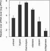
Effect of various proteases on virus replication in VeroE6 cells. VeroE6 cells in 24-well plates were infected as described in Fig. 4 and cultured in the presence of trypsin (62.5 μg/ml), thermolysin (12.5 μg/ml), elastase (125 μg/ml), papain (0.037 unit/ml), or collagenase (200 μg/ml). At 20 h after infection, the amounts of mRNA9 were measured by real-time PCR. Relative viral mRNA level is displayed by virus infectivity (pfu) calculated from the calibration line.

References

    1. Ksiazek, T .G., Erdman, D., Goldsmith, C., Zaki, S. R., Peret, T., Emery, S., Tong, S., Urbani, C., Comer, J. A., Lim, W., et al. (2003) N. Engl. J. Med. 348, 1953-1966. - PubMed
    1. Drosten, C., Gunther, S., Preiser, W., Van Der Werf, S., Brodt, H. R., Becker, S., Rabenau, H., Panning, M., Kolesnikowa, L., Fouchier, R. A., et al. (2003) N. Engl. J. Med. 348, 1967-1976. - PubMed
    1. Marra, M. A., Jones, S. J., Astell, C. R., Holt, R. A., Brooks-Wilson, A., Buttefield, Y. S., Khattra, J., Asano, J. K., Barber, S. A., Chan, S. Y., et al. (2003) Science 300, 1399-1404. - PubMed
    1. Rota, P. A., Oberste, M. S., Monroe, S. S., Nix, W. A., Campagnoli, R., Icenogle, J. P., Penaranda, S., Bankamp, B., Maher, K., Chen, M. H., et al. (2003) Science 300, 1394-1399. - PubMed
    1. The Chinese SARS Molecular Epidemiology Consortium (2004) Science 303, 1666-1669. - PubMed

Publication types

MeSH terms

LinkOut - more resources