

Longitudinal volumetric MRI change and rate of cognitive decline
- PMID: 16116117
- PMCID: PMC1820871
- DOI: 10.1212/01.wnl.0000172913.88973.0d
Longitudinal volumetric MRI change and rate of cognitive decline
Abstract
Objective: To examine how baseline and change of volumetric MRI relate to cognitive decline in older individuals.
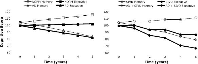
Background: Memory is associated with hippocampal integrity, whereas executive function has been linked to impaired frontal lobe function. Previous studies have shown that hippocampal and cortical atrophy are more strongly related to cognition than are measures of subcortical cerebrovascular disease (CVD). The authors hypothesized that memory (MEM) decline would be related to change in hippocampal volume (HC), whereas decline in executive function (EXEC) would be related to change of cortical gray matter volume (CGM) and measures of subcortical CVD.
Methods: Subjects from a multicenter study (n = 103) included cognitively normal, mildly impaired, and demented cases with and without subcortical lacunes. All had longitudinal cognitive evaluation (mean = 4.8 years) and two or more MRI scans at least one year apart (mean = 3.4 years). MRI measures included HC, CGM, total lacune volume (LAC), and white matter hyperintensity volume (WMH). Random effects modeling of longitudinal data assessed effects of MRI baseline and MRI change on baseline and change of psychometrically matched measures of MEM and EXEC.
Results: Change in MEM was related to HC baseline and HC change. Change in EXEC was related to baseline CGM and to change in CGM, HC, and LAC. Results were unchanged when demented cases were excluded. WMH was not associated with change in MEM or EXEC independent of HC, CGM, and LAC.
Conclusion: Hippocampal volume was the primary determinant of memory decline, whereas executive function (EXEC) decline was related to multiple brain components. Results support a hypothesis that MEM decline is strongly influenced by Alzheimer disease (AD), whereas EXEC decline may be complexly determined by cerebrovascular disease and AD.
Figures
References
-
- Lobo A, Launer LJ, Fratiglioni L, et al. Neurologic Diseases in the Elderly Research Group Prevalence of dementia and major subtypes in Europe: a collaborative study of population based cohorts. Neurology. 2000;54(11 suppl 5):S4–S9. - PubMed
-
- Nyenhuis DL, Gorelick PB. Vascular dementia: a contemporary review of epidemiology, diagnosis, prevention, and treatment. J Am Geriatr Soc. 1998;46:1437–1448. - PubMed
-
- Lopez OL, Kuller LH, Fitzpatrick A, Ives D, Becker JT, Beauchamp N. Evaluation of dementia in the cardiovascular health cognition study. Neuroepidemiology. 2003;22:1–12. - PubMed
-
- Knopman DS, Parisi JE, Boeve BF, et al. Vascular dementia in a population based autopsy study. Arch Neurol. 2003;60:569–575. - PubMed
-
- Visser PJ, Scheltens P, Verhey FR, et al. Medial temporal lobe atrophy and memory dysfunction as predictors for dementia in subjects with mild cognitive impairment. J Neurol. 1999;246:477–485. - PubMed
Publication types
MeSH terms
Grants and funding
LinkOut - more resources
Full Text Sources
Medical