. 2005 Sep 1;175(5):3092-3101.
doi: 10.4049/jimmunol.175.5.3092.

NKT cell stimulation with glycolipid antigen in vivo: costimulation-dependent expansion, Bim-dependent contraction, and hyporesponsiveness to further antigenic challenge

Affiliations

NKT cell stimulation with glycolipid antigen in vivo: costimulation-dependent expansion, Bim-dependent contraction, and hyporesponsiveness to further antigenic challenge

Adam P Uldrich et al. J Immunol. .

Abstract

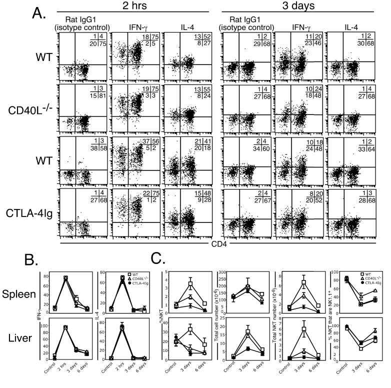
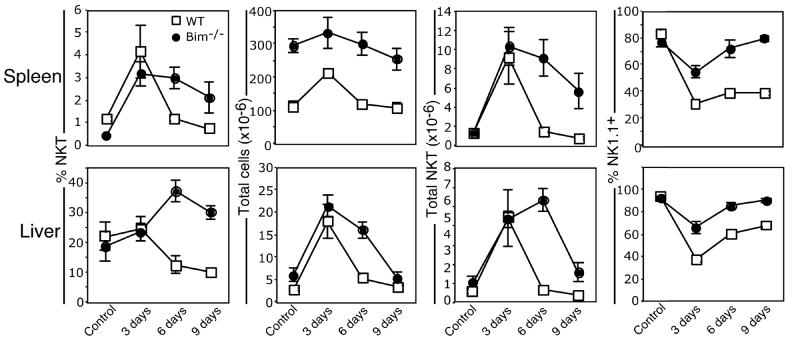
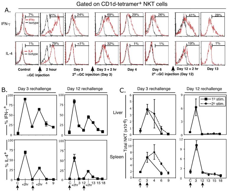
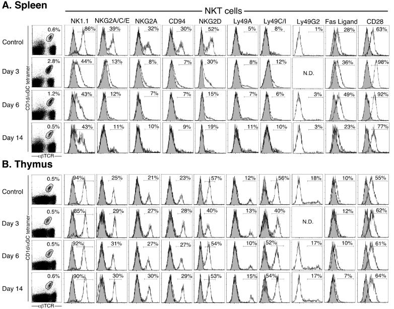
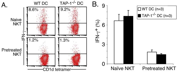
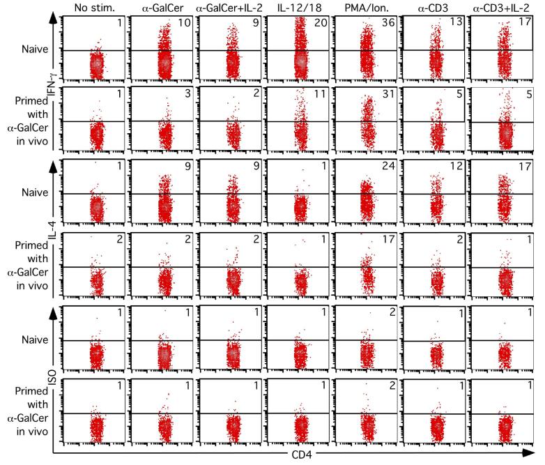
Activation of NKT cells using the glycolipid alpha-galactosylceramide (alpha-GalCer) has availed many investigations into their immunoregulatory and therapeutic potential. However, it remains unclear how they respond to stimulation in vivo, which costimulatory pathways are important, and what factors (e.g., Ag availability and activation-induced cell death) limit their response. We have explored these questions in the context of an in vivo model of NKT cell dynamics spanning activation, population expansion, and subsequent contraction. Neither the B7/CD28 nor the CD40/CD40L costimulatory pathway was necessary for cytokine production by activated NKT cells, either early (2 h) or late (3 days) after initial stimulation, but both pathways were necessary for normal proliferative expansion of NKT cells in vivo. The proapoptotic Bcl-2 family member Bim was necessary for normal contraction of the NKT cell population between days 3-9 after stimulation, suggesting that the pool size is regulated by apoptotic death, similar to that of conventional T cells. Ag availability was not the limiting factor for NKT cell expansion in vivo, and a second alpha-GalCer injection induced a very blunted response, whereby cytokine production was reduced and further expansion did not occur. This appeared to be a form of anergy that was intrinsic to NKT cells and was not associated with inhibitory NK receptor signaling. Furthermore, NKT cells from mice pre-challenged with alpha-GalCer in vivo showed little cytokine production and reduced proliferation in vitro. In summary, this study significantly enhances our understanding of how NKT cells respond to primary and secondary antigenic challenge in vivo.


Figures

Figure 1.
Figure 1.
CD40 and CD28 signalling are both essential for NKT cell expansion but dispensable for cytokine production. Mice (WT, CD40L-/- and CTLA4-Ig transgenic) were injected i.p. with 2 μg α-GalCer and examined between 2 hrs and 6 days later. Liver and spleen-derived lymphocytes were isolated and cultured for 2 hrs in GolgiStop, surface-labelled with CD1d/α-GalCer tetramer or anti-αβTCR, CD4 or NK1.1 mAb, and then fixed and permeabilized for intracellular staining with antibodies to IL-4 or IFN-γ. NKT cells were electronically gated and analysed for their expression of IFN-γ or IL-4 (A and B). A. displays representative flow cytometry data from each group at each time point. Quadrants were set based on staining with the rat IgG1 isotype control antibody. B. Pooled data of IFN-γ and IL-4 production and is representative of (WT: C = control (PBS injected) 8 mice; 2 hr = 8 mice; 3 days = 6 mice; 6 days = 3 mice; CD40L-/-: C = control (PBS injected) 2 mice; 2 hr = 2 mice; 3 days = 2 mice; 6 days = 2 mice; CTLA-4IgTg: C = control (PBS injected) = 5 mice; 2 hr = 5 mice; 3 days = 6 mice; 6 days = 3 mice; between 1-3 experiments/strain/time-point. Control (PBS injected) mice were harvested at each time-point to control for unstimulated NKT cell status. (C) Graphs from left to right show the percentage CD1d/α-GalCer-tetramer-binding NKT cells, total number of lymphocytes per organ, total number of NKT cells per organ and percentage of NKT cells that were NK1.1+. For B and C, the top row shows spleen and bottom row shows liver. Error bars represent the standard error of the mean.
Figure 2.
Figure 2.
Contraction of the NKT cell pool is bim-dependent Mice (WT and bim-/-) were injected i.p. with 2 μg α-GalCer and examined between 3 and 9 days later. Liver and spleen-derived lymphocytes were isolated and stained with CD1d/α-GalCer tetramer, anti-αβTCR and NK1.1 mAbs and analysed for the percentage of CD1d/α-GalCer tetramer-binding αβTCR+ NKT cells, total lymphocyte number and total NKT cell number in both the spleen (top row) and liver (bottom row). Data are derived from two independent experiments with a total of n = (WT: = 5 mice; 3 days = 5 mice. 6 days = 5 mice; 9 days = 5 mice; Bim-/-: C (Control, PBS injected) = 5 mice, 3 days = 5 mice. 6 days = 6 mice; 9 days = 5 mice). Control (PBS injected) mice were harvested at each time-point to control for unstimulated NKT cell status. Error bars represent the standard error of the mean.
Figure 3.
Figure 3.
The NKT cell response to α-GalCer re-challenge in vivo. Mice (WT) were injected i.p. with 2 μg α-GalCer and re-injected on day 3 or day12 after primary challenge. Liver and spleen-derived lymphocytes were isolated and cultured for 2 hrs in Brefeldin-A, surface-labelled with CD1d/α-GalCer tetramer or anti-αβTCR, CD4 or NK1.1 mAbs, and then fixed and permeabilized for intracellular staining with mAbs to IL-4 or IFN-γ. CD1d/α-GalCer tetramer αβTCR+ NKT cells were gated and analysed for their expression of IFN-γ or IL-4, as shown in (A) with representative data from liver NKT cells after a 3 day or 12 day rechallenge experiment. Accumulated cytokine data from day 3 and day 12 rechallenge experiments are shown in B. Total NKT cell numbers at each time-point are shown in C. Data were derived from 3-5 mice per group. Control (PBS injected) mice were harvested at each timepoint to control for unstimulated NKT cell status. Error bars represent the standard error of the mean. Arrows indicate the time of α-GalCer injection.
Figure 4
Figure 4
Modulation of cell surface receptors on NKT cells following α-GalCer stimulation in vivo. Mice (WT) were injected i.p. with 2 μg α-GalCer on day 0, and examined 3, 6, or 14 days later. Spleen and thymus-derived lymphocytes were isolated and stained with CD1d/α-GalCer tetramer plus anti-αβTCR (first column), and NKT cells were electronically gated as shown and examined for expression of NK1.1, NKG2A/C/D/E, CD94, Ly49A/C/G2/I, Fas-Ligand (CD178) and CD28 (open histograms), overlayed onto relevant isotype controls (greyed histograms). Controls (no injection), n = 3 mice; day 3, n = 2 mice; day 6, n = 3 mice; day 14, n = 3 mice.
Figure 5
Figure 5
NKT cell anergy is autonomous and independent of NK inhibitory receptor ligation. Mice (WT) were injected i.p. with 2 μg α-GalCer or PBS, and spleen-derived lymphocytes were isolated 7 days later, B cell depleted using magnetic beads, and NKT cells sorted based on CD1d/α-GalCer tetramer-binding and αβTCR expression. CD11c+ DCs were also sorted from WT and TAP-1-/- mice. Purified NKT cells from naïve or α-GalCer-pretreated mice were cocultured with WT or TAP-1-/- DCs, at a ratio of 5:1 for 12 hin vitro ± 200 ng/ml α-GalCer, and subsequently stained for anti-IFN-γ-PE, or rat IgG1-PE isotype control. (A) Representative profiles of IFN-γ production by gated CD1d/α-GalCer tetramer+ αβTCR+ NKT cells and (B) Graph depicts mean and standard error for each group. IFN-γ staining regions were set based on the isotype control, and data is representative of 3 mice per group.
Figure 6.
Figure 6.
Previously challenged NKT cells show reduced in vitro proliferation following re-challenge with α-GalCer. Spleen-derived lymphocytes were labelled with CFSE and cultured for 1 day with α-GalCer with or without 50 units/ml IL-2, washed and the culture continued for an additional 2.5 days without α-GalCer (to allow T cell receptor re-expression), but with IL-2 replenished in appropriate cultures. Following culture, NKT cells were stained with CD1d/α-GalCer tetramer and anti-αβTCR mAb and examined for evidence of cell division as indicated by dilution of CFSE-labeling. The percentage of cells having undergone one or more divisions is shown, as well as the geometric mean fluorescence intensity (MFI) for the entire NKT cell population. This data represents results from 1 of 4 similar experiments carried out between 2 weeks and 2 months after primary in vivo α-GalCer stimulation.
Figure 7.
Figure 7.
Impaired in vitro cytokine production by NKT cells from mice previously challenged with α-GalCer. Mice were injected with 2 μg α-GalCer i.p. and killed 1 month later. Liver-derived lymphocytes were isolated and cultured for 6 hours with no further stimulation, 200ng/mL α-GalCer ± 50 units/mL IL-2, 100ng/mL each IL-12 and IL-18, 10ng/mL PMA and 3μM ionomycin, or 10μg/mL plate-bound anti-CD3 ± 50 units/ml IL-2. GolgiStop was added for the final 5 hours of culture before cells were surface-stained with the CD1d/α-GalCer tetramer and anti-CD4, and then fixed and permeabilized for intracellular staining with anti-IFN-γ, anti-IL-4 or rat IgG1 isotype control intracellular mAb. CD1d/α-GalCer tetramer+ CD4+ cells were selected by electronic gating and analysed for their expression of IFN-γ, for specific staining by the isotype control. Data is representative of 4 mice per group and this is experiment is representative of 4 experiments carried out between 2 weeks and 2 months after primary in vivo α-GalCer treatment.

References

    1. Godfrey DI, MacDonald HR, Kronenberg M, Smyth MJ, Van Kaer L. NKT cells: what's in a name? Nat Rev Immunol. 2004;4:231. - PubMed
    1. Godfrey DI, Kronenberg M. Going both ways: immune regulation via CD1d-dependent NKT cells. J Clin Invest. 2004;114:1379. - PMC - PubMed
    1. Brigl M, Brenner MB. CD1: Antigen Presentation and T Cell Function. Annu Rev Immunol. 2004;22:817. - PubMed
    1. Kronenberg M. Toward an understanding of NKT cell biology: Progress and Paradoxes. Annu Rev Immunol. 2005;23:877. - PubMed
    1. Hayakawa Y, Godfrey DI, Smyth MJ. Alpha-galactosylceramide: potential immunomodulatory activity and future application. Curr Med Chem. 2004;11:241. - PubMed

Publication types