. 2005 Sep;37(9):958-63.
doi: 10.1038/ng1626. Epub 2005 Aug 21.

A human ortholog of archaeal DNA repair protein Hef is defective in Fanconi anemia complementation group M

Affiliations

A human ortholog of archaeal DNA repair protein Hef is defective in Fanconi anemia complementation group M

Amom Ruhikanta Meetei et al. Nat Genet. 2005 Sep.

Abstract

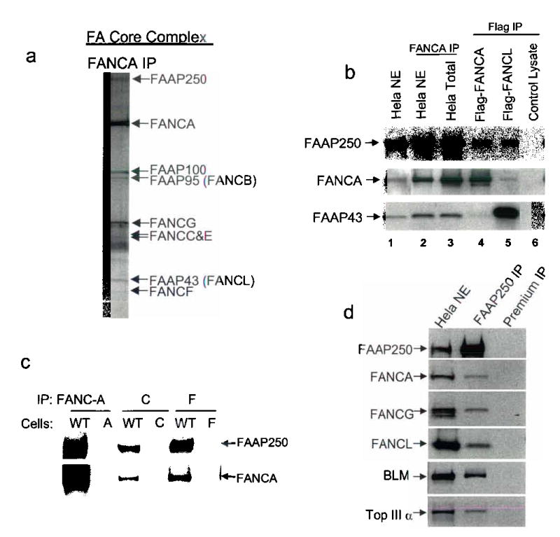
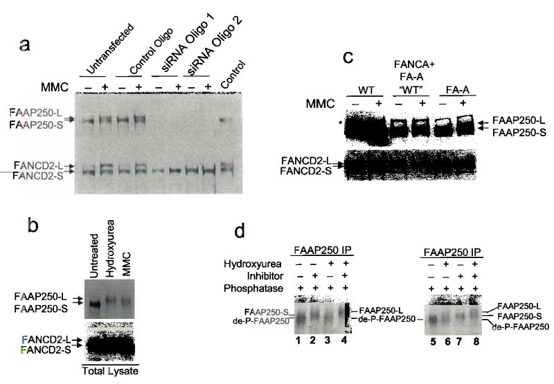
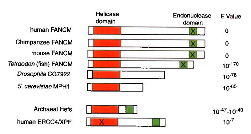
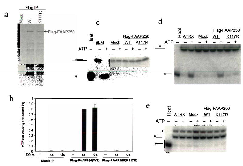
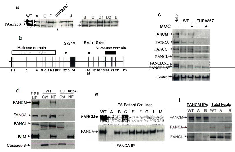
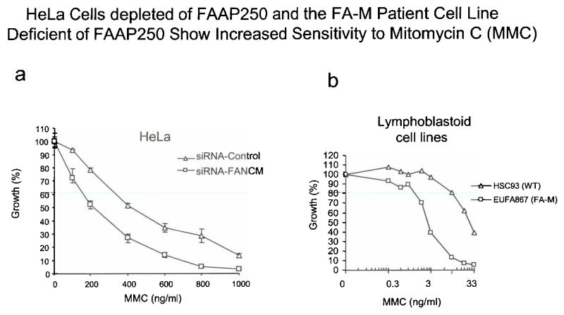
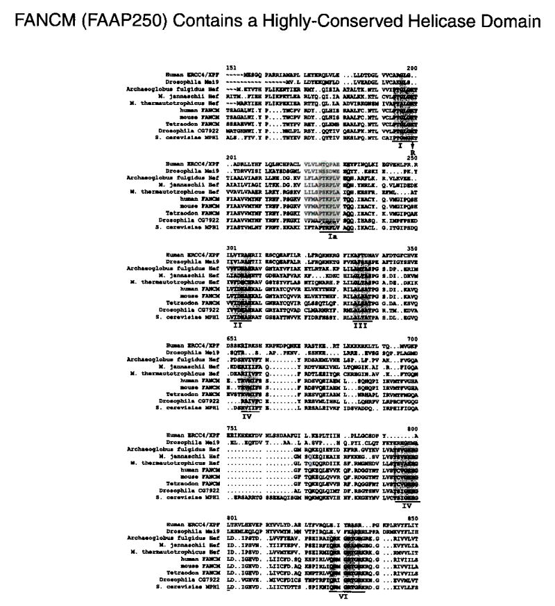
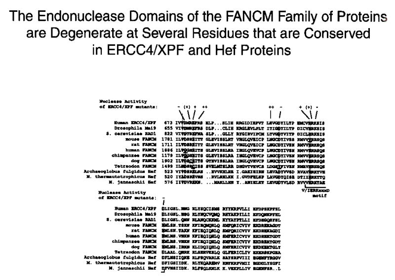
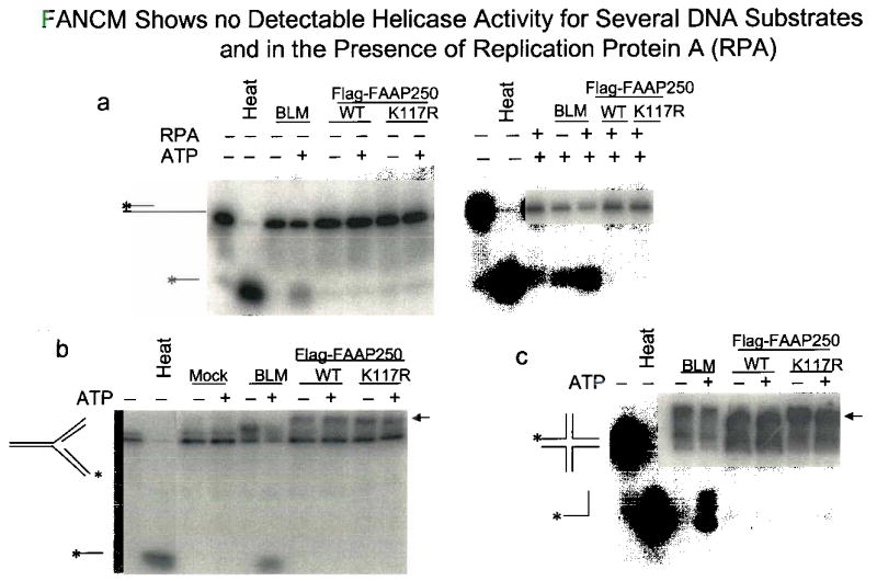
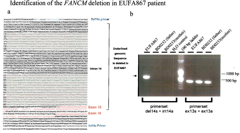
Fanconi anemia is a genetic disease characterized by genomic instability and cancer predisposition. Nine genes involved in Fanconi anemia have been identified; their products participate in a DNA damage-response network involving BRCA1 and BRCA2 (refs. 2,3). We previously purified a Fanconi anemia core complex containing the FANCL ubiquitin ligase and six other Fanconi anemia-associated proteins. Each protein in this complex is essential for monoubiquitination of FANCD2, a key reaction in the Fanconi anemia DNA damage-response pathway. Here we show that another component of this complex, FAAP250, is mutant in individuals with Fanconi anemia of a new complementation group (FA-M). FAAP250 or FANCM has sequence similarity to known DNA-repair proteins, including archaeal Hef, yeast MPH1 and human ERCC4 or XPF. FANCM can dissociate DNA triplex, possibly owing to its ability to translocate on duplex DNA. FANCM is essential for monoubiquitination of FANCD2 and becomes hyperphosphorylated in response to DNA damage. Our data suggest an evolutionary link between Fanconi anemia-associated proteins and DNA repair; FANCM may act as an engine that translocates the Fanconi anemia core complex along DNA.


Conflict of interest statement

Competing Interests Statement The authors declare that they have no competing financial interests.

Figures

Figure 1
Figure 1. FAAP250 is an integral component of the FA core complex
(a) A silver stained SDS gel showing the immunopurified FA core complex. We have previously shown that this complex containing 5 known FA proteins (FANCA, -C, -E, -F and -G) and four novel components, which are termed FAAPs for FANCA -associated polypeptides,. We demonstrated that two FAAPs are also FA proteins: FAAP95 is FANCB, and FAAP43 is FANCL, a novel E3 ubiquitin ligase. (b) Immunoblotting to show the presence of FAAP250 in polypeptides obtained from immunoprecipitation (IP) using antibodies against FANCA from HeLa nuclear extract and whole cell extract. FAAP250 was also co-immunoprecipitated by Flag antibody from HEK293 cells stably expressing Flag-FANCA and Flag-FANCL. As a control, FAAP250 was not co-immunoprecipitated by a mock immunoprecipitation using HEK293 cells that do not express the Flag-tagged FA proteins (lane 6). (c) Immunoblotting to show co-IP of FAAP250 with FANCA, FANCC and FANCF from cell lysate of a normal individual (WT), but not from those of FA patients defective in the corresponding FA genes, as indicated. (d) Immunoblotting shows that multiple FA core complex proteins were co-immunoprecipited by a FAAP250 antibody. A preimmune serum was used in a control immunoprecipitation. The presence of BLM and Topo IIIa, which stably associate with the FA core proteins in the BRAFT complex, was also shown.
Fig. 2
Fig. 2. FAAP250 is required for FANCD2 monoubiquitination and is hyperphosphorylated in response to DNA damage
(a) Immunoblotting shows that HeLa cells depleted of FAAP250 by two different siRNA oligos have defective FANCD2 monoubiquitination. HeLa nuclear extract was used as a control. We noticed that a new form of FAAP250 with slower mobility, marked as FAAP250-L (long form), was induced in cells treated with MMC or other genotoxic agents. FAAP250 from unstressed cells was marked as FAAP250-S (short form). (b) Immunoblotting shows that FAAP250 alters its gel mobility when HEK293 cells were treated with hydroxyurea and MMC. FANCD2 monoubiquitination was used as a control. (c) Immunoblotting shows induction of FAAP250-L in lymphoblastoid cells from a normal individual (WT), an FA-A patient and the same patient cell line complemented by expression of exogenous FANCA. (d) Immunoblotting shows that both FAAP250-S and FAAP250-L increased their mobility upon treatment by lamda protein phosphatase. Notably, the de-phosphorylated FAAP250 (de-P-FAAP250) has a faster mobility than either FAAP250-S or FAAP250-L, suggesting both forms of FAAP250 are phosphorylated. We found that our immunoisolated FAAP250 may contain some contaminating phosphatases, which, in the presence of the phosphatase buffer, can dephosphorylate FAAP250 to some degree even without the addition of the exogenous phosphatase. To circumvent this problem, we used the phosphatase inhibitor-treated FAAP250 fraction as the un-dephosphorylated control.
Fig. 3
Fig. 3. FAAP250 (FANCM) contains Potential DNA-Metabolizing Domains and is homologous to DNA repair proteins in Archea and yeast
A schematic presentation of DNA-metabolizing domains in FAAP250 (named FANCM here; see text) and its sequence homologues. The helicase and endonuclease domains are marked by red and green, respectively, and their sequence alignments are shown in Supplementary Fig. 2 and 3. The domains marked by the “X” are predicted to be non-functional based on degeneracy of their sequences compared to the consensus. The helicase domains of ERCC4/XPF proteins have been suggested to be degenerate and perhaps nonfunctional ,. The E values were obtained by searching the NCBI database with the BLASTP program and the human FANCM as the query sequence. The homology between MPH1 and a partial EST sequence of FAAP250 has been described.
Fig. 4
Fig. 4. FAAP250 has an ATP-dependent DNA translocase activity
(a) A silver-stained SDS-gel showing the immunoisolated Flag-FAAP250 of wildtype and K117R mutant from HEK293-derived cells stably expressing these proteins. Polypeptides immunoisolated from HEK293 cells that do not express the Flag-tagged proteins are shown as a control (Mock). (b) A histogram shows that Flag-FAAP250 has DNA-stimulated ATPase activity, whereas a point mutation within the conserved helicase motif I (K117R) inactivates such activity. The double-stranded (ds) and single-stranded (ss) DNA are indicated. (c) An autoradiograph showing that Flag-FAAP250 exihibits no detectable helicase activity in a double-strand displacement assay. BLM helicase-associated complexes (BLM) were used as a positive control. The DNA substrate has 13-nucleotide single-stranded tails on both sides of a 20-bp double-stranded region, so that it should be able to detect activity of helicases of either 5′ to 3′ or 3′ to 5′ directions. (d) Autoradiograph showing that Flag-FAAP250 displays a triple-helix unwinding activity, whereas its ATPase mutant (K117R) lacks such activity. A chromatin-remodeling complex, ATRX complex, was used as a positive control. The substrate consists of a 40-base homopyrimidine oligonucleotide located centrally on a 190 bp double-stranded DNA. (e) Autoradiograph showing that Flag-FAAP250 does not displace a blunt triple helix. The substrate is the same as in (d), except that the duplex DNA is the same size (40 bp) as the homopyrimidine oligonucleotide, to create the blunt triplex. The band with faster mobility is from the designed blunt triplex substrate. The band with slower mobility (marked with an arrowhead) is from the precursor of the substrate.
Fig. 5
Fig. 5. FAAP250 is mutated in Fanconi anemia patients and is essential for FA core complex assembly and function
(a) Immunoblotting shows that FAAP250 is absent in a FA patient of a new complementation group, EUFA867. The phenotype of this patient is typical of FA, with increased chromosomal breakage on treatment of cells with MMC and DEB, progressive bone marrow failure, and characteristic bilateral radial anomalies. A nonspecific polypeptide crossreactive with FAAP250 antibody (marked with an asterisk) could be used as a loading control. (b) A schematic representation of the genomic structure of FAAP250 and the mutations (S724X and exon 15 deletion) detected EUFA867 and her sibling, who also has FA. The nonsense mutation was also detected in the mother of the two patients. (c) Immunoblotting shows that in EUFA867 cell extract, the levels of FANCA and FANCG are strongly reduced, whereas that of FANCL is moderately reduced, when compared to the extract from a normal individual (WT). In addition, monoubiquitinated FANCD2 (FANCD2-L) was not detectable in the extract of EUFA867 cells. (d) Immunoblotting shows defective nuclear localization of FANCA and FANCL in EUFA867 cells. HeLa nuclear extract (NE) was included as a control. BLM and caspase 3 were used as markers for nuclear (NE) and cytoplasmic (cyt) extracts, respectively. (e) Immunoprecipitation-coupled immunoblotting shows that FANCM failed to co-immunoprecipitate with FANCA from extracts of FA patient cells lacking various core complex proteins, as indicated on the top. (f) Immunoprecipitation coupled-immunoblotting shows reduced co-immunoprecipitation of FANCA and FANCL by an FANCM antibody in FA-A and FA-B cell extracts.
Supplementary Fig. 1
Legend: (a) A graph showing that HeLa cells depleted of FAAP250 (FANCM) by siRNA have increased MMC sensitivity compared to cells treated with a control siRNA oligo. (b) A graph showing that the FA-M patient-derived lymphoblastoid cell line (EUFA867) deficient of FAAP250(FANCM) has increased sensitivity to MMC compared to a cell line derived from a normal individual (WT). It was noticed that the increased MMC sensitivity is about 2-fold for HeLa cells, lower than that of FA-M cells (about 10-fold). This difference could be due to the limitation of siRNA technique that fails to completely deplete FAAP250. There is a difference in the two procedures: in experiment (a), cells were treated with a high dose of MMC for 2 hrs, whereas in experiment (b), cells were continuously exposed to a low dose of MMC for a period of 3 doubling times (between 3–14 days).
Supplementary Fig. 2
Legend: Sequence alignment of predicted helicase domains of FANCM and its sequence homologs. The seven motifs present in all superfamily II helicases are underlined, and the conserved residues are highlighted. The arrow indicates the conserved lysine (K117) in motif I that has been replaced by arginine in Flag-FANCIvl ATPase mutant. ERCC4/XPF and Mei9 (the Drosophila homolog of ERCC4/XPF) are included in the alignment.
Supplementary Fig. 3
Legend: The alignment of the C-terminal endonuclease domains of FANCM (FAAP250) proteins with those in ERCC4/XPF and Hefs. The residues that are absolutely conserved in ERCC4/XPF, Mus81 and Hef families of proteins are highlighted, and those that are degenerate in FANCM protein are underlined. A summary of the endonuclease activity of various ERCC4/XPF point mutants is shown at the top. The different signs are: “++” for wildtype activity; “+” for no detectable activity in the presence of Mg++, but residue activity in the presence of Mn++; “(+)”, little or no activity; “−”, no activity. A conserved motif found in many nucleases is indicated by a bracket.
Supplementary Fig. 4
Legend: (a) An audioradiograph shows that the presence of a 40-nucleotide 3′ single-stranded DNA tail in the substrate and the addition of RPA (100 nM) fail to activate the potential helicase activity of Flag-FANCM. These conditions can activate the helicase activity of MPH1, the yeast orthologue of FANCM. The presence of a 40-nucleotide 5′ single-stranded DNA tail in the substrate also fails to activate the helicase activity of FANCM (data not shown), (b) and (c) Autoradiographs show that FANCM cannot unwind two structured DNA substrates that can be resolved by Hef, the archaeal ortholgue of FANCM. The arrow indicates a possible complex between structured DNA and Flag-FAAP250.
Supplementary Fig. 5
Legend: (a) Sequence of the F.4AO/genomic DNA surrounding the deleted region (underlined). Exons are marked with red. The two PCR primers used amplify the region deleted in the patient in (B) are indicated in blue. (b) BD0232 and BD0233 are lymphoblastoid cell lines from the father and mother of EUFA867. The PCR product from the wild type allele is non-detectable, due to the large size of this region which cannot be efficiently amplified under the current condition. Only the PCR product from the deleted allelle is detected (marked with an arrow). The PCR on exon 13 was performed as a control and shows the integrity of the genomic DNA.

Comment in

Similar articles

Cited by

References

    1. Joenje H, Patel KJ. The emerging genetic and molecular basis of fanconi anaemia. Nat Rev Genet. 2001;2:446–59. - PubMed
    1. Garcia-Higuera I, et al. Interaction of the Fanconi anemia proteins and BRCA1 in a common pathway. Mol Cell. 2001;7:249–62. - PubMed
    1. Howlett NG, et al. Biallelic Inactivation of BRCA2 in Fanconi Anemia. Science. 2002;13:13. - PubMed
    1. Meetei AR, et al. A multiprotein nuclear complex connects Fanconi anemia and bloom syndrome. Mol Cell Biol. 2003;23:3417–3426. - PMC - PubMed
    1. Meetei AR, et al. A novel ubiquitin ligase is deficient in Fanconi anemia. Nat Genet. 2003;35:165–70. - PubMed

Publication types

MeSH terms

Associated data