Comparative Study
. 2005 Dec 1;392(Pt 2):271-81.
doi: 10.1042/BJ20050873.

Comparison of the SUMO1 and ubiquitin conjugation pathways during the inhibition of proteasome activity with evidence of SUMO1 recycling

Affiliations
Comparative Study

Comparison of the SUMO1 and ubiquitin conjugation pathways during the inhibition of proteasome activity with evidence of SUMO1 recycling

Daniel Bailey et al. Biochem J. .

Abstract

To investigate potential interplay between the SUMO1 (small ubiquitin-related modifier-1) and ubiquitin pathways of post-translational protein modification, we examined aspects of their localization and conjugation status during proteasome inhibition. Our results indicate that these pathways converge upon the discrete sub-nuclear domains known as PML (promyelocytic leukaemia protein) NBs (nuclear bodies). Proteasome inhibition generated an increased number of PML bodies, without any obvious increase in size. Using a cell line that constitutively expresses an epitope-tagged version of SUMO1, which was incorporated into high-molecular-mass conjugates, we observed SUMO1 accumulating in clusters around a subset of the NBs. Nuclear ubiquitin was initially observed in numerous speckles and foci, which bore no relationship to PML NBs in the absence of proteasome inhibition. However, during proteasome inhibition, total ubiquitin-conjugated species increased in the cell, as judged by Western blotting. Concomitantly the number of nuclear ubiquitin clusters decreased, and were almost quantitatively associated with the PML NBs, co-localizing with the SUMO-conjugated pool. Proteasome inhibition depleted the pool of free SUMO1 in the cell. Reversal of proteasome inhibition in the presence or absence of protein synthesis demonstrated that free SUMO1 was regenerated from the conjugated pool. The results indicate that a significant fraction of the free SUMO1 pool could be accounted for by recycling from the conjugated pool and indeed it may be that, as for ubiquitin, SUMO1 needs to be removed from conjugated species prior to processing by the proteasome. Taken together with other recent reports on the proteasome and PML NBs, these results suggest that the PML NBs may play an important role in integrating these pathways.


Figures

Figure 1
Figure 1. Effects of proteasome inhibition by MG132 on endogenous PML and SUMO1
Hep2 cells were treated for 7 h with either DMSO (control) or 10 μM MG132. Cells were subsequently fixed with methanol and immunofluorescence was performed to detect endogenous PML (a and d) using an anti-PML polyclonal antibody (DB#75) and endogenous SUMO1 (b and e) using anti-PIC1 antibody. Corresponding merged panels are shown (c and f); arrows indicate examples of co-localization of SUMO1 and PML at the NBs.
Figure 2
Figure 2. Localization of SUMO1 in a cell line constitutively expressing epitope-tagged SUMO1
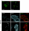
A Hep2-SUMO cell line expressing a myc epitope-tagged SUMO1 protein (hmSUMO1) was fixed and immunofluorescence staining was performed to detect hmSUMO1, using an anti-myc antibody (9E10), or with a polyclonal antibody to detect PML. Expression of hmSUMO1 was mainly nuclear diffuse, although some dense staining nuclear domains were also observed. The arrows in the separate images indicate dense staining areas, enriched for SUMO1 that co-localized with PML (see merge image).
Figure 3
Figure 3. Effect of proteasome inhibition (MG132) on PML and exogenous SUMO1
(a) Hep2-SUMO cells were treated for up to 7 h with DMSO (Control) or 10 μM MG132. Cells were subsequently fixed with methanol and immunofluorescence was performed to detect epitope-tagged SUMO1 (red) using anti-myc antibody, and endogenous PML (green) using an anti-PML polyclonal antibody (#75). Arrows indicate representative NBs showing recruitment of hmSUMO1. (b) Similar results were observed with Hep2-SUMO cells treated for 7 h with either 20 μM PSI or 50 μM lactacystin and processed in a similar manner. MG132-treated and control cells are shown for comparative purposes.
Figure 4
Figure 4. Analysis of a non-conjugatable variant of SUMO1
(a) Hep2-SUMO cells were separately transfected with mock, wild-type HA-SUMO1 or the HA-SUMO NC variant. Cells were subsequently harvested by cell lysis, extracts separated by electrophoresis, and immunoblotting performed. Separations were probed for HA-SUMO1 using an antibody to the HA-epitope tag. (b) Hep2-SUMO-NC cells were separately transfected with wild-type HA-SUMO1 (ac and gi) or (HA-SUMO-NC (df and jl). Cells were treated with DMSO (control) or 10 μM MG132 for 5 h, prior to fixation and staining with anti-HA antibody for the transfected SUMO1, or with antibody 9E10 for the integrated myc-tagged SUMO1. The wild-type HA-SUMO localized with the standard SUMO1 profile (a versus b), whereas the HA-SUMO-NC variant was diffuse (d versus e) throughout the cytoplasm and nucleus. Upon MG132 treatment the wild-type HA-SUMO was recruited to SUMO1 domains (g versus h, short arrows), whereas the HA-SUMO-NC remained diffuse and was not recruited to SUMO1 domains (j versus k, long arrows). Respective merge images are shown in c, f, i and l.
Figure 5
Figure 5. Localization of SUMO1 in relation to conjugated ubiquitin
Hep2-SUMO cells treated with DMSO (control) or 10 μM MG132 for 7 h were fixed with methanol and immunofluorescence analysis was performed. (a) Conjugated ubiquitin was detected with anti-ubiquitin FK2 monoclonal antibody (green), and PML with anti-PML #75 polyclonal antibody (red). Arrows indicate PML domains. (b) Hep2-SUMO cells co-stained for conjugated ubiquitin with anti-ubiquitin FK2 (green) and SUMO1 with anti-PIC1 antibody (red). Short arrows indicate representative domains with SUMO1 and ubiquitin co-localization, whilst long arrows indicate accumulation of cytoplasmic ubiquitin.
Figure 6
Figure 6. Western blot analysis of SUMO1 modification and ubiquitination during MG132 treatment
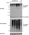
Hep2-SUMO cells treated with 10 μM MG132 for 7 h were harvested by cell lysis. Cell extracts were separated by electrophoresis and immunoblotting performed. SDS/10–20%-PAGE gel separations were probed for hmSUMO1 using an anti-myc (9E10) antibody. SDS/3–8%-PAGE gel separations were probed for conjugated ubiquitin using anti-ubiquitin FK2. Equivalent samples were also probed with an anti-actin polyclonal antibody as a control for loading.
Figure 7
Figure 7. Effect of proteasome release on SUMO1 conjugation
(a) Schematic of the time course experiment. Hep2-SUMO cells treated with DMSO (control) or 20 μM MG132 for 7 h were washed and media replaced±cycloheximide at 100 μg/ml. Cells were incubated for up to 6 h prior to harvesting, then separated by electrophoresis and immunoblotting performed. (b) Extracts were probed for SUMO1 (using an anti-myc antibody). Equivalent samples were also probed with an anti-actin polyclonal antibody as a control for loading.
Figure 8
Figure 8. A speculative model on the relationship between the accumulation of ubiquitinated and SUMO1-modified proteins at the NBs
This proposal summarizes one potential model for how the accumulation of SUMO1 conjugates and the appearance of ubiquitin conjugates at the NB may be regulated. In this proposal SUMO1- modified proteins (a) dynamically traffic through the nuclear body (b) wherein SUMO-specific proteases participate in the removal of SUMO1, in a manner which is possibly co-ordinated with the ubiquitination pathway. SUMO1 may be exchanged for ubiquitin either at the same or different lysines and the proteins targeted for degradation by the proteasome (c). MG132 treatment blocks the proteasome resulting in the stacking up of proteins at and around the NB (d).

References

    1. Gill G. SUMO and ubiquitin in the nucleus: different functions, similar mechanisms? Genes Dev. 2004;18:2046–2059. - PubMed
    1. Weissman A. M. Themes and variations on ubiquitylation. Nat. Rev. Mol. Cell Biol. 2001;2:169–178. - PubMed
    1. Okura T., Gong L., Kamitani T., Wada T., Okura I., Wei C. F., Chang H. M., Yeh E. T. Protection against Fas/APO-1- and tumor necrosis factor-mediated cell death by a novel protein, sentrin. J. Immunol. 1996;157:4277–4281. - PubMed
    1. Boddy M. N., Howe K., Etkin L. D., Solomon E., Freemont P. S. PIC 1, a novel ubiquitin-like protein which interacts with the PML component of a multiprotein complex that is disrupted in acute promyelocytic leukaemia. Oncogene. 1996;13:971–982. - PubMed
    1. Matunis M. J., Coutavas E., Blobel G. A novel ubiquitin-like modification modulates the partitioning of the Ran-GTPase-activating protein RanGAP1 between the cytosol and the nuclear pore complex. J. Cell Biol. 1996;135:1457–1470. - PMC - PubMed

Publication types

MeSH terms