. 2005 Sep 6;102(36):12771-6.
doi: 10.1073/pnas.0505758102. Epub 2005 Aug 23.

Effects of the tumor inhibitory triterpenoid avicin G on cell integrity, cytokinesis, and protein ubiquitination in fission yeast

Affiliations

Effects of the tumor inhibitory triterpenoid avicin G on cell integrity, cytokinesis, and protein ubiquitination in fission yeast

Jordan U Gutterman et al. Proc Natl Acad Sci U S A. .

Abstract

Avicins comprise a class of triterpenoid compounds that exhibit tumor inhibitory activity. Here we show that avicin G is inhibitory to growth of the fission yeast Schizosaccharomyces pombe. S. pombe cells treated with a lethal concentration of avicin G (20 microM) exhibited a shrunken morphology, indicating that avicin G adversely affects cell integrity. Cells treated with a sublethal concentration of avicin G (6.5 microM) exhibited a strong cytokinesis-defective phenotype (multiseptated cells), as well as cell morphology defects. These phenotypes bear resemblance to those resulting from loss of Rho1 GTPase function in S. pombe. Indeed, Rho1-deficient S. pombe cells were strongly hypersensitive to avicin G, suggesting that the compound may perturb Rho1-dependent processes. Consistent with previously observed effects in human Jurkat T cells, avicin G treatment resulted in hyperaccumulation of ubiquitinated proteins in S. pombe cells. Interestingly, proteasome-defective S. pombe mutants were not markedly hypersensitive to avicin G, whereas an anaphase-promoting complex (mitotic ubiquitin ligase) mutant exhibited avicin G resistance, suggesting that the increase in levels of ubiquitinated proteins resulting from avicin G treatment may be due to increased protein ubiquitination, rather than inhibition of 26S proteasome activity. Mutants defective in the cAMP/PKA pathway also exhibited resistance to avicin G. Our results suggest that S. pombe will be a useful model organism for elucidating molecular targets of avicin G and serve as a guide to clinical application where dysfunctional aspects of Rho and/or ubiquitination function have been demonstrated as in cancer, fibrosis, and inflammation.


Figures

Fig. 1.
Fig. 1.
Avicin G inhibits S. pombe cell growth. (A) Wild-type S. pombe cells (105) were spread on a YEAU plate. Five-microliter volumes of a 1:4 dilution series of avicin G (dissolved in water) were spotted onto the cell lawn (0.1–25 μg of drug), which were incubated for 3 days at 30°C. Five microliters of water was spotted on the plate as a control (C). Growth inhibition (circular areas devoid of cell growth) was observed where 1.6 μg, 6.3 μg, and 25 μg amounts of the drug were spotted. (B) Wild-type S. pombe cells were incubated in YEAU (control) or YEAU containing the indicated concentrations of avicin G and monitored for growth for 12 h. Cell densities (cells per ml) were determined microscopically by using a hemacytometer. (C) Wild-type S. pombe cells were incubated in YEAU to low density, then diluted with fresh YEAU to a density of 2.5 × 10⁵ cells per ml. Avicin G was added to half the culture to a final concentration of 20 μg/ml, and an equal volume of water was added to the remaining culture. At 1, 2, 4, and 8 h, 10-ml portions of each culture were pelleted by centrifugation, washed twice with YEAU, and resuspended in YEAU. Cell densities were determined by using a hemacytometer, and 2,000 cells from each sample were spread onto YEAU plates. After 3 days, colonies were counted. The graph shows the frequency of cell viability (based on colony-forming units) of untreated and avicin G-treated cultures at the indicated time points. Duplicate samples were cultured for each time point.
Fig. 2.
Fig. 2.
Microscopic analysis of avicin G-treated S. pombe cells. Photomicrographs of wild-type S. pombe cells incubated in YEAU (A) or YEAU containing 40 μg/ml avicin G (B) for 10 h. The majority of avicin G-treated cells were shrunken in appearance. (C) Photomicrograph of wild-type S. pombe cells treated with a sublethal concentration of avicin G (10 μg/ml) for 10 h. A high frequency of cells contained multiple septa. (D) Fluorescence photomicrograph of DAPI-stained S. pombe cells treated with 10 μg/ml avicin G for 10 h. Three multiseptated cells are shown, the compartments of which each contained a single nucleus.
Fig. 3.
Fig. 3.
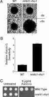
Rho1-deficient S. pombe cells are hypersensitive to avicin G. (A) Wild-type and nmt41-rho1 cells were overlaid onto EMM (Upper) or EMM plus 0.1 μM thiamine (EMMT) (Lower) as described for Fig. 1. Avicin G (25 μg) was spotted onto each cell lawn, and the plates were incubated for 3 days at 30°C. On EMMT plates, but not EMM plates, the area of avicin G-induced growth inhibition was significantly greater for nmt41-rho1 lawns than the wild-type cell lawns. (B) Relative avicin G sensitivity of wild-type and nmt41-rho1 cells based on areas of growth inhibition from two separate drug sensitivity tests of the type shown in A. Avicin G sensitivity of wild-type cells was normalized to a value of 1. (C) Serial dilutions (1:5) of wild-type and nmt41-rho1 cells were spotted onto EMMT or EMMT containing 8 μg/ml avicin G and incubated for 5 days at 30°C. Wild-type cells grew on the avicin G plate, whereas nmt41-rho1 cells did not.
Fig. 4.
Fig. 4.
Relative avicin G sensitivities of stress-response-defective S. pombe mutants. (A) Wild-type and spc1Δ S. pombe strains were overlaid onto YEAU plates as described in Fig. 1. Avicin G (25 μg) was then spotted onto the respective cell lawns, and the plates were incubated at 30°C for 3 days. The relative avicin G sensitivity of each strain, based on measurements of areas of avicin G-induced growth inhibition, was then determined, with wild-type cells being normalized to a value of 1. (B) Serial dilutions (1:5) of wild-type, cyr1Δ, pka1Δ, and plb1Δ cells were spotted onto YEAU or YEAU containing 16 μg/ml avicin G and incubated for 3 days at 30°C. cyr1Δ, pka1Δ, and plb1Δ cells, but not wild type, grew on the avicin-G-containing plate.
Fig. 5.
Fig. 5.
Avicin G causes hyperaccumulation of ubiquitinated proteins in S. pombe cells. Wild-type S. pombe cells were incubated in YEAU containing 20 μg/ml avicin G for the time indicated (h), then processed for immunoblot analysis of ubiquitinated proteins. An increase in the levels of ubiquitinated proteins was detected after 1.5 h of avicin G treatment.
Fig. 6.
Fig. 6.
Effects of avicin G on the growth of 26S proteasome and APC-defective S. pombe mutants. Wild-type, mts2-1 (mts2), mts3-1 (mts3), and nuc2-663 cells were spread onto a YEAU plates as described for Fig. 1. Avicin G (25 μg) was then spotted onto the respective cell lawns, and the plates were incubated at 26°C for 5 days. The relative avicin G sensitivity of each strain, based on measurements of areas of avicin G-induced growth inhibition, was then determined, with wild-type cells being normalized to a value of 1. (B) Serial dilutions (1:5) of wild-type and nuc2-663 cells were spotted onto YEAU or YEAU containing 16 μg/ml avicin G and incubated for 5 days at 26°C. nuc2-663 cells, but not wild-type cells, grew on the avicin G-containing plate.

References

    1. Haridas, V., Arntzen, C. J. & Gutterman, J. U. (2001) Proc. Natl. Acad. Sci. USA 98, 11557–11562. - PMC - PubMed
    1. Haridas, V., Higuchi, M., Jayatilake, G. S., Bailey, D., Mujoo, K., Blake, M. E., Arntzen, C. J. & Gutterman, J. U. (2001) Proc. Natl. Acad. Sci. USA 98, 5821–5826. - PMC - PubMed
    1. Haridas, V., Hanausek, M., Nishimura, G., Soehnge, H., Gaikwad, A., Narog, M., Spears, E., Zoltaszek, R., Walaszek, Z. & Gutterman, J. U. (2004) J. Clin. Invest. 113, 65–73. - PMC - PubMed
    1. Hanausek, M., Ganesh, P., Walaszek, Z., Arntzen, C. J., Slaga, T. J. & Gutterman, J. U. (2001) Proc. Natl. Acad. Sci. USA 98, 11551–11556. - PMC - PubMed
    1. Mujoo, K., Haridas, V., Hoffmann, J. J., Wachter, G. A., Hutter, L. K., Lu, Y., Blake, M. E., Jayatilake, G. S., Bailey, D., Mills, G. B. & Gutterman, J. U. (2001) Cancer Res. 61, 5486–5490. - PubMed

Publication types

MeSH terms

LinkOut - more resources