. 2005 Dec 15;106(13):4076-85.
doi: 10.1182/blood-2005-06-2204. Epub 2005 Aug 23.

Differential roles of microtubule assembly and sliding in proplatelet formation by megakaryocytes

Affiliations

Differential roles of microtubule assembly and sliding in proplatelet formation by megakaryocytes

Sunita R Patel et al. Blood. .

Abstract

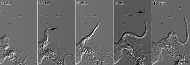
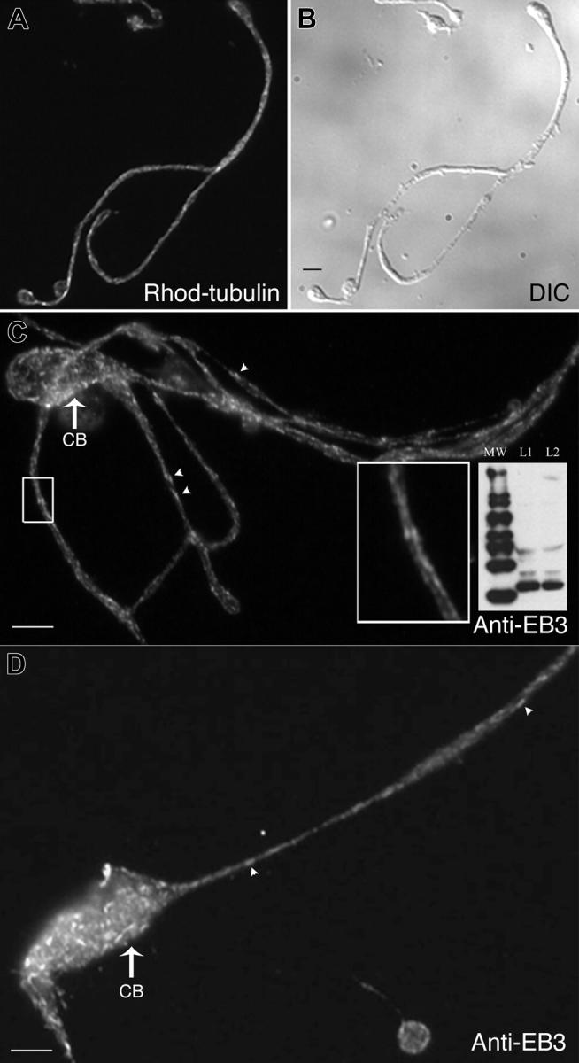
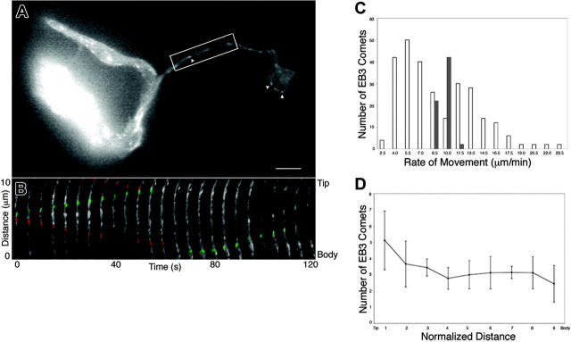
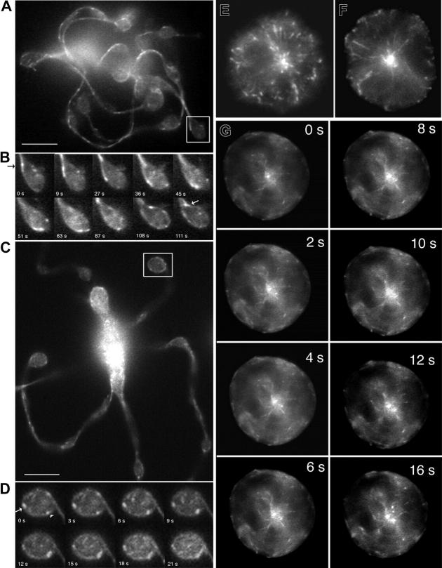
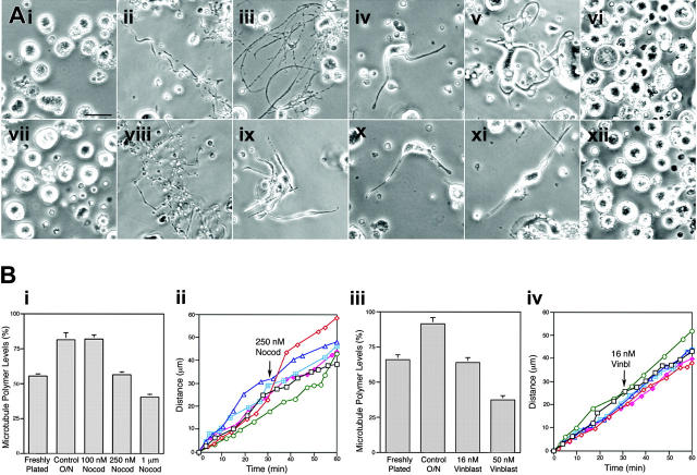
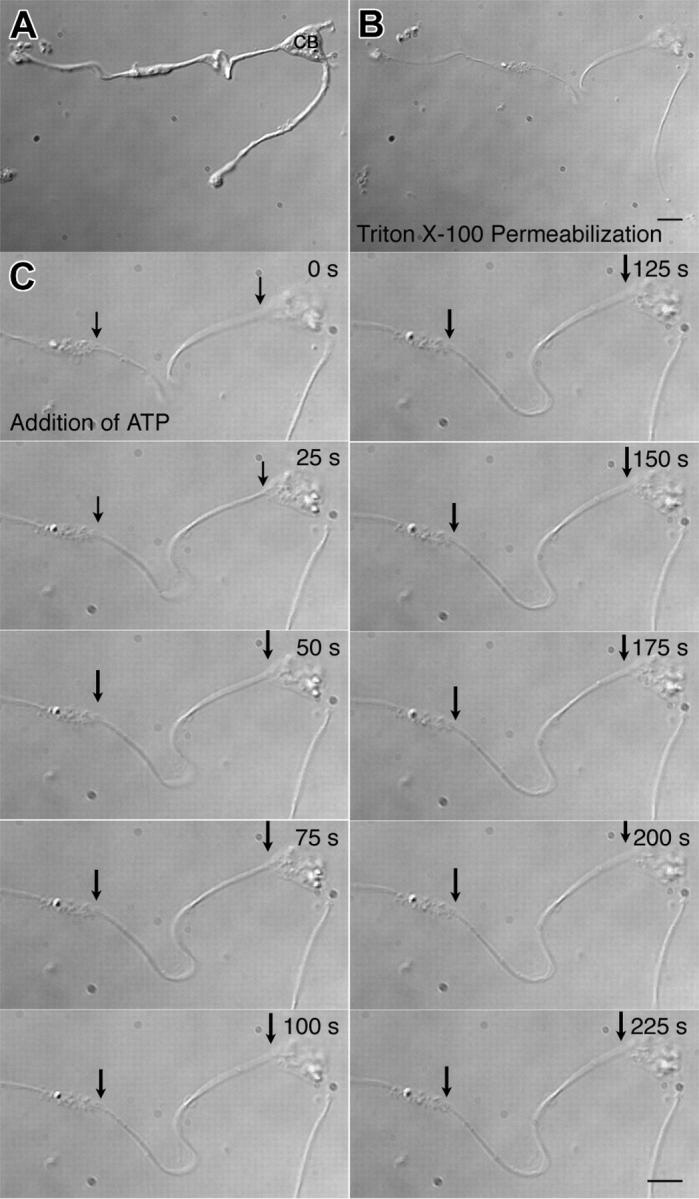
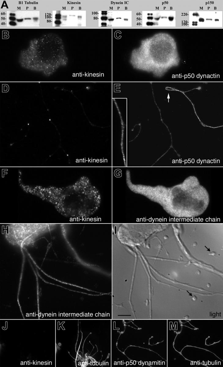
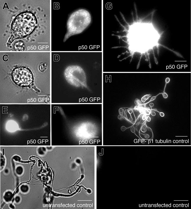
Megakaryocytes are terminally differentiated cells that, in their final hours, convert their cytoplasm into long, branched proplatelets, which remodel into blood platelets. Proplatelets elongate at an average rate of 0.85 microm/min in a microtubule-dependent process. Addition of rhodamine-tubulin to permeabilized proplatelets, immunofluorescence microscopy of the microtubule plus-end marker end-binding protein 3 (EB3), and fluorescence time-lapse microscopy of EB3-green fluorescent protein (GFP)-expressing megakaryocytes reveal that microtubules, organized as bipolar arrays, continuously polymerize throughout the proplatelet. In immature megakaryocytes lacking proplatelets, microtubule plus-ends initiate and grow by centrosomal nucleation at rates of 8.9 to 12.3 microm/min. In contrast, plus-end growth rates of microtubules within proplatelets are highly variable (1.5-23.5 microm/min) and are both slower and faster than those seen in immature cells. Despite the continuous assembly of microtubules, proplatelets continue to elongate when net microtubule assembly is arrested. One alternative mechanism for force generation is microtubule sliding. Triton X-100-permeabilized proplatelets containing dynein and its regulatory complex, dynactin, but not kinesin, elongate with the addition of adenosine triphosphate (ATP) at a rate of 0.65 microm/min. Retroviral expression in megakaryocytes of dynamitin (p50), which disrupts dynactin-dynein function, inhibits proplatelet elongation. We conclude that while continuous polymerization of microtubules is necessary to support the enlarging proplatelet mass, the sliding of overlapping microtubules is a vital component of proplatelet elongation.


Figures

Figure 1.
Figure 1.
Video-enhanced differential-interference-contrast microscopy showing a representative proplatelet elongating from a mouse megakaryocyte. By 3 minutes (arrow), the initial broad pseudopodia has converted into a proplatelet process that continues to lengthen and reduce its diameter at the proplatelet tip (arrow at 9 minutes). Proplatelets elongate at an average rate of 0.85 ± 0.24 μm/min (n = 77). Bar, 5 μm. See Movie S1.
Figure 2.
Figure 2.
Localization of microtubule plus-ends in proplatelets. (A-B) Megakaryocytes extending proplatelets were permeabilized with Triton X-100 and incubated with TRITC (tetramethylrhodamine-isothiocyanate)–tubulin as described in “Materials and methods.” (A) Fluorescence micrograph showing a permeabilized proplatelet after incubation with rhodamine-tubulin for 4 minutes. TRITC-tubulin incorporates into specific foci along the length of the proplatelet. Bar, 4 μm. (B) Differential-interference-contrast micrograph of cell labeled in panel A. (C-D) Anti-EB3 immunofluorescent labeling of a proplatelet-containing megakaryocyte. EB3 staining (arrowheads label cometlike dashes) is dispersed along proplatelets and is abundant in the cell bodies (CB) but is not found in a radial pattern (compare with preproplatelet megakaryocytes in Figure 5). The boxed region in panel C shows a high-magnification view of the comets. Bars, 4 μm. (C, inset) The immunoblot shows that anti-EB3 antibodies recognize a 36-kDa polypeptide in both (L1) preproplatelet megakaryocytes and (L2) proplatelet-containing megakaryocytes. MW lane indicates molecular weight protein ladder.
Figure 3.
Figure 3.
EB3-GFP movements in proplatelet-producing megakaryocytes. (A) First frame from a time-lapse movie of a live megakaryocyte expressing EB3-GFP (Movie S2). The cell body is at the left of the micrograph. EB3-GFP labels growing microtubule plus-ends in a characteristic “comet” staining pattern (arrowheads) that has a bright front and dim tail. Moving comets are found along the proplatelets as well as in the megakaryocyte cell body. The scale bar is 5 μm. (B) Kymograph of the boxed region in panel A. Images are every 5 seconds. EB3-GFP comets undergo bidirectional movements in proplatelets. Some EB3-GFP comets move tipward and are highlighted in green; others that move toward the cell body are highlighted in red. (C) Comparison of the velocity distribution of comets moving in proplatelets (white bars) with those emanating from the centrosome of preproplatelet megakaryocytes (dark bars). The average rate of comet movement in the preproplatelet megakaryocytes was 10.2 ± 0.77 μm/min and the rates of movement were tightly grouped (8.9-12.3 μm/min). EB3 movements in proplatelets, however, are apparently bimodal with distinct populations moving slower and faster than those of the preproplatelet megakaryocytes. (D) EB3-GFP comets were distributed throughout the proplatelet. Proplatelets were divided into 10 segments (tip = 0, cell body = 9) and the number of GFP-EB3 comets in each was determined. The length of the evaluated proplatelet was 28 μm. Error bars indicate standard deviation
Figure 4.
Figure 4.
EB3-GFP comet movements in living megakaryocytes. (A-D) EB3-GFP comet movements within the tips of proplatelets. (A) First frame of a time-lapse sequence shown in Movie S3. (C) First frame of a time-lapse sequence shown in Movie S4. (B,D) Kymograph of EB3 around proplatelet tips. In the first example, EB3-GFP comets enter the proplatelet tip, circle its periphery, and then reenter the shaft of the proplatelet. Images are every 5 seconds. In the second example, EB3-GFP comets move around the periphery of the tip in both directions (arrowhead highlights clockwise movement; arrow highlights counterclockwise movement). Images are every 2 seconds. Comets also move into the tip on microtubules that end abruptly. The scale bar represents 5 μm. (E-G) Location and dynamics of microtubule plus-ends in representative preproplatelet megakaryocytes. (E-F) Anti-EB3 immunofluorescence of preproplatelet-megakaryocytes. Cells at this stage of maturation have a radial array of microtubules that emanates from the centrosome. EB3-GFP comets concentrate near the centrosome and are on the plus-ends of microtubules that radiate outward. Field width, 20 μm. (G) Dynamics of EB3-GFP comets in a megakaryocyte lacking proplatelets. This sequence of images shows EB3-GFP to concentrate on the plus-ends of microtubules as they grow from the centrosome (Movie S4). Translocations of EB3-GFP in the cell cortex, parallel to the plasma membrane, are also apparent. Elapsed time is in seconds.
Figure 5.
Figure 5.
Effect of microtubule assembly inhibitors on proplatelet elaboration. (Ai-x) Phase-contrast micrographs of mouse megakaryocytes grown for 20 hours in the (ii, viii) absence or (iii-vi, ix-xii) presence of microtubule inhibitors. (i-vii) Freshly plated megakaryocytes lack proplatelet extensions. Megakaryocytes cultured in the presence of (iii) 100 nM, (iv-v) 250 nM, and (vi) 1 μm nocodazole, or (ix-xi) 16 nM and (xii) 50 nM vinblastine. In control cultures, the cells become decorated with long proplatelets in 20 hours. Proplatelets were elaborated normally when the cells were cultured in (iii) 100 nM nocodazole and some proplatelets were found on cells cultured in either (iv-v) 250 nM nocodazole or (ix-xi) 16 nM vinblastine, although extensions are shorter and thicker compared with those elaborated in the absence of the inhibitors. Proplatelet formation is completely inhibited by (vi) 1 μm nocodazole or (xii) 50 nM vinblastine. Scale bar, 25 μm. (Bi) Effect of increasing concentrations of nocodazole on tubulin polymer levels in megakaryocytes. The graph compares the percentage of tubulin polymerized into microtubules in freshly plated megakaryocytes lacking proplatelets (∼ 55%) to megakaryocytes plated for 20 hours in the absence (control) or presence of 100 nM, 250 nM, and 1 μm nocodazole. Culturing of megakaryocytes for 20 hours in the absence or presence of 100 nM nocodazole resulted in an increase of total tubulin polymer to approximately 85% (a 25.9% increase from freshly plated cells). Tubulin polymer levels in megakaryocytes cultured in the presence of 250 nM nocodazole remained stable relative to the initial value, showing that 250 nM nocodazole acts as a kinetic stabilizer of microtubules. The tubulin polymer content of cultured megakaryocytes was decreased to approximately 40% by 1 μm nocodazole. (Bii) Proplatelet elongation is unaffected by 250 nM nocodazole. The rate of elongation was studied in 6 proplatelets before and after treatment with 250 nM nocodazole. Nocodazole was added after 30 minutes (arrow). (Biii) Effect of increasing concentrations of vinblastine on tubulin polymer levels in megakaryocytes. The graph compares the percentage of total tubulin polymerized into microtubules in freshly plated megakaryocytes lacking proplatelets (65%) to megakaryocytes plated for 20 hours in the absence (control) or presence of 16 nM and 50 nM vinblastine. Culturing of megakaryocytes for 20 hours increased the total tubulin polymer content of cells to approximately 90%. Megakaryocytes cultured in the presence of 16 nM and 50 nM vinblastine were unable to increase their polymer content or had diminished tubulin polymer contents, respectively. (Biv) Proplatelet elongation is unaffected by 16 nM vinblastine. The rate of elongation was studied in 6 proplatelets before and after treatment with 16 nM vinblastine, added at time 30 minutes (arrow). Error bars indicate standard deviations.
Figure 6.
Figure 6.
Activation of elongation in a Triton X-100–permeabilized proplatelet by ATP. Changes in proplatelet length after the addition of ATP were monitored by microscopy. (A) A proplatelet viewed with DIC optics just before detergent permeabilization. Two proplatelets can be observed extending from the cell body (CB) of a megakaryocyte. (B) Treatment with 0.5% Triton X-100, followed by washing in a microtubule-stabilizing buffer, preserves the general structure of the proplatelet. (C) Time-lapse sequence after the addition of 1 mM ATP. ATP causes the proplatelet residue to increase its contour length and individual microtubules to splay apart from the bundle. Note the increase in distance between the cell body (right arrow) and the swelling that was attached to the substrate (left arrow). The rate of elongation in this example is approximately 0.7 μm/min. The lengthening of the proplatelet slows after 125 seconds. Scale bar, 5 μm. See Movie S6.
Figure 7.
Figure 7.
Immunolocalization of cytoplasmic dynein, dynactin, and kinesin in megakaryocytes. (A) Characterization of antibodies by immunoblotting. Whole-cell protein extracts from mouse megakaryocytes (M), mouse platelets (P), and rat brain (B) were displayed by SDS-PAGE, transferred to PVDF, and immunoblotted with antibodies against β1-tubulin, kinesin, dynein intermediate chain, p50 dynactin, and p150Glued. Double immunofluorescence microscopy of megakaryocytes using antibodies to (B,D,F) antikinesin and (C,E) either anti-p50 dynactin or (G) anti–dynein intermediate chain antibodies. Immunofluorescence images of preproplatelet megakaryocytes and proplatelet-containing megakaryocytes. (B,F) Kinesin antibodies stain vesicle-like particles within megakaryocytes and (D) along the shafts of proplatelets. (C) p50 dynactin and dynein intermediate chain antibodies diffusely stain the megakaryocyte cytoplasm. Proplatelets are intensely stained (E, inset) along their length with anti-p50 dynactin and (H) anti–dynein intermediate chain antibodies. (H) Comparative immunofluorescent and (I) DIC images of a proplatelet-containing megakaryocyte and mouse platelets (arrows) stained with anti–dynein intermediate chain antibodies. Proplatelets stain robustly with anti–dynein intermediate chain antibody. In contrast, staining of platelets seeded onto the coverslip is highly reduced. Scale bar, 5 μm. Dynactin remains associated with the Triton X-100–insoluble megakaryocyte cytoskeleton (J-M). Megakaryocytes were extracted with 0.5% Triton X-100 in a microtubule-stabilizing buffer before fixation, as described in “Materials and methods.” The cells were then immunostained using (J) kinesin and (L) p50 dynamitin and (K,M) counterstained with α-tubulin antibodies to visualize proplatelet microtubules. Kinesin immunoreactivity was removed by the detergent treatment, suggesting that kinesin is not associated with the microtubules. In contrast, the p50 dynamitin immunoreactivity is preserved.
Figure 8.
Figure 8.
Role of dynactin in proplatelet elongation. (A-J) Effects of p50 dynamitin expression on proplatelet elongation. (A,C) Differential-interference-contrast and (B,D-G) fluorescence images of p50-GFP–expressing cells exhibiting a range of elongation distortions, compared with the unperturbed elongation of representative megakaryocytes (H) retrovirally directed to express GFP–β1-tubulin and (I-J) uninfected, nonexpressing cells. All images were acquired by fluorescence and DIC microscopy. Scale bars all represent 10 μm.

References

    1. Choi ES, Nichol JL, Hokom MM, Hornkohl AC, Hunt P. Platelets generated in vitro from proplatelet-displaying human megakaryocytes are functional. Blood. 1995;85: 402-413. - PubMed
    1. Cramer EM, Norol F, Guichard J, et al. Ultrastructure of platelet formation by human megakaryocytes cultured with the Mpl ligand. Blood. 1997;89: 2336-2346. - PubMed
    1. Leven RM, Yee MK. Megakaryocyte morphogenesis stimulated in vitro by whole or partially fractionated thrombocytopenic plasma: a model system for the study of platelet formation. Blood. 1987;69: 1046-1052. - PubMed
    1. Radley J, Rogerson J, Ellis S, Hasthorpe S. Megakaryocyte maturation in long-term marrow culture. Exp Hematol. 1991;19: 1075-1078. - PubMed
    1. Radley JM, Scurfield G. The mechanism of platelet release. Blood. 1980;56: 996-999. - PubMed

Publication types