. 2005 Dec 15;106(13):4093-101.
doi: 10.1182/blood-2005-02-0671. Epub 2005 Aug 23.

Bone marrow dysfunction in mice lacking the cytokine receptor gp130 in endothelial cells

Affiliations

Bone marrow dysfunction in mice lacking the cytokine receptor gp130 in endothelial cells

Longbiao Yao et al. Blood. .

Abstract

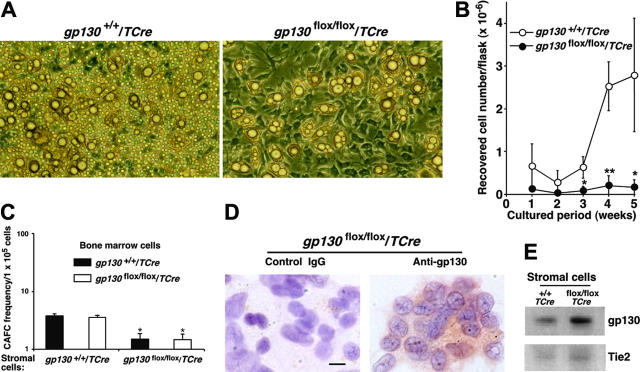
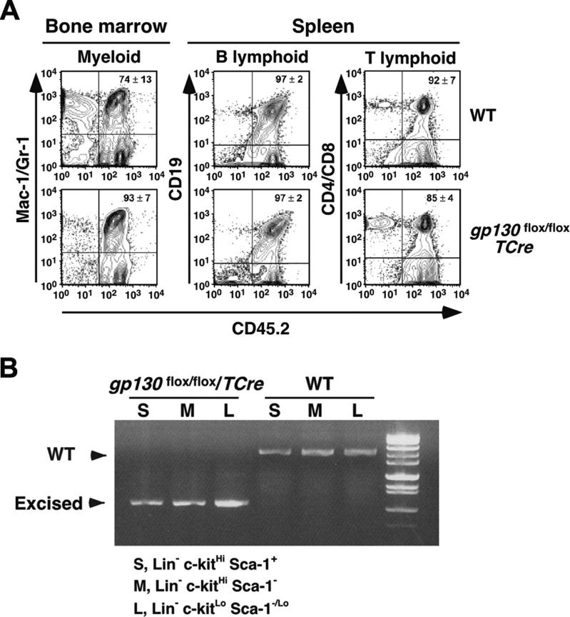
In vitro studies suggest that bone marrow endothelial cells contribute to multilineage hematopoiesis, but this function has not been studied in vivo. We used a Cre/loxP-mediated recombination to produce mice that lacked the cytokine receptor subunit gp130 in hematopoietic and endothelial cells. Although normal at birth, the mice developed bone marrow dysfunction that was accompanied by splenomegaly caused by extramedullary hematopoiesis. The hypocellular marrow contained myeloerythroid progenitors and functional repopulating stem cells. However, long-term bone marrow cultures produced few hematopoietic cells despite continued expression of gp130 in most stromal cells. Transplanting gp130-deficient bone marrow into irradiated wild-type mice conferred normal hematopoiesis, whereas transplanting wild-type bone marrow into irradiated gp130-deficient mice did not cure the hematopoietic defects. These data provide evidence that gp130 expression in the bone marrow microenvironment, most likely in endothelial cells, makes an important contribution to hematopoiesis.


Figures

Figure 1.
Figure 1.
Generation of gp130flox/flox/TCre mice. (A) Schematic diagram of portions of the WT gp130 allele, the gp130flox/flox allele, and the gp130flox/flox allele after Cre-mediated excision of the floxed exon. Exon numbers are shown. The loxP sites are depicted as large arrowheads adjacent to the introduced XhoI restriction sites. Small arrowheads indicate the sites of PCR primers that were used to amplify each allele. (B) Genotype analysis. gp130 flox/+/TCre mice were bred with gp130flox/+ mice. PCR products of genomic DNA from the offspring were resolved on agarose gels before or after digestion with XhoI. Arrows indicate the products corresponding to the WT allele, the floxed allele, and the floxed allele after Cre-mediated excision of the floxed exon.
Figure 2.
Figure 2.
Specific deletion of gp130 in hematopoietic and endothelial cells of gp130flox/flox/TCre mice. (A) Immunoblots of tissue lysates probed with antibodies to gp130 or to the control protein Stat3. (B) Immunoblots of sorted CD45+ splenic T cells expressing CD4 or CD8 and of cultured lung endothelial cells (ECs) probed with antibodies to gp130 or Stat3. Cells were pooled from 3 mice from each genotype. (C) Confocal immunofluorescence microscopy of lung sections stained with antibodies to the endothelial-cell marker PECAM-1 (green) and to gp130 (red). Yellow staining indicates colocalization of PECAM-1 and gp130 in the endothelial cells of gp130+/+/TCre mice. Only green staining for PECAM-1 is present in the endothelial cells of gp130flox/flox/TCre mice. Both sections exhibit red staining for gp130 in nonendothelial cells. Scale bar represents 20 μm. Objective, × 40/0.75 NA. (D) Immunoperoxidase staining of bone marrow sections with antibodies to VWF or to gp130. Red staining for VWF is present in megakaryocytes (arrows) and sinusoidal endothelial cells (arrowheads) of both gp130+/+/TCre mice and gp130flox/flox/TCre mice. Staining for gp130 is present in megakaryocytes, a subset of other hematopoietic cells, and endothelial cells of gp130+/+/TCre mice. By contrast, there is no staining for gp130 in hematopoietic or endothelial cells of gp130flox/flox/TCre mice. Scale bar represents 50 μm. Data are representative of 3 to 4 independent experiments with 12-week-old mice. Objective, × 40/0.75 NA.
Figure 3.
Figure 3.
Shortened survival and defective hematopoiesis in gp130flox/flox/TCre mice. (A) Shortened survival of gp130flox/flox/TCre mice. (B) Peripheral-blood smear of a 16-week-old gp130flox/flox/TCre mouse exhibits polychromasia and anisopoikilocytosis of erythrocytes. Scale bar represents 20 μm. Objective, × 63/1.4 NA. (C) Splenomegaly in a 12-week-old gp130flox/flox/TCre mouse. Scale bar represents 1 cm. (D) Spleen of a 12-week-old gp130flox/flox/TCre mouse exhibits extramedullary hematopoiesis with abundant megakaryocytes. Bone marrow of a 16-week-old gp130flox/flox/TCre mouse exhibits normal cellularity in the endosteal niche (EN) adjacent to bone. In contrast, the vascular niche (VN) is markedly hypocellular, except for retention of normal numbers of megakaryocytes. Scale bar represents 50 μm. Objective, × 20/0.5 NA. (E) Quantification of blood cells of B-cell, T-cell, myeloid, and erythroid lineage expressing the indicated markers in bone marrow (BM), spleen, and thymus of gp130+/+/TCre and gp130flox/flox/TCre mice. Data represent the mean ± SD of 5 mice (age 12 weeks) in each genotype. *P < .05 and **P < .01 compared with gp130+/ +/TCre.
Figure 4.
Figure 4.
gp130flox/flox/TCre bone marrow retains progenitor cells. (A) Quantification of the indicated myeloerythroid colonies that developed from bone marrow or spleen cells suspended in methylcellulose. Data represent the mean ± SD of triplicate determinations and are representative of 3 independent experiments. *P < .001 compared with gp130+/+/TCre. (B) To determine the relative proportions of hematopoietic stem/progenitor cells, Lin- bone marrow cells were stained with PE–anti-Sca-1 and APC–anti-c-kit and then were analyzed by flow cytometry. The c-kitHiSca-1+, c-kitHiSca-1-, and c-kitLoSca-1-/Lo populations are gated with open boxes. The percentage of cells in each population is shown. (C) Quantification of the indicated myeloerythroid colonies that developed from 200 sorted Lin-c-kitHiSca-1+ cells suspended in methylcellulose. Data represent the mean ± SD of triplicate determinations from cells pooled from 5 mice per genotype and are representative of 2 independent experiments.
Figure 5.
Figure 5.
gp130flox/flox/TCre bone marrow does not produce hematopoietic cells in long-term cultures. (A) Photomicrographs of long-term bone marrow cultures. The stromal-cell monolayer appears normal in the gp130flox/flox/Cre culture, but there are markedly reduced numbers of nonadherent hematopoietic cells. Magnification, × 100. (B) Quantification of myeloid cells produced in long-term bone marrow cultures. Data represent the mean ± SD of triplicate determinations and are representative of at least 3 independent experiments. *P < .05 and **P < .01 compared with gp130+/+/TCre. (C) Quantification of CAFC frequency in long-term bone marrow cultures plated on irradiated stromal cells of the indicated genotype. *P < .05 compared with bone marrow cells plated on gp130+/+/TCre stromal cells. (D) Immunocytochemistry of stromal cells in gp130flox/flox/TCre cultures probed with control or anti-gp130 antibodies. Scale bar represents 10 μm. (E) Immunoblots of lysates of stromal cells from long-term bone marrow cultures probed with antibodies to gp130 or Tie2. Data are representative of 3 independent experiments.
Figure 6.
Figure 6.
Serial transplantation of gp130-deficient bone marrow reconstitutes hematopoiesis in irradiated mice. (A) Bone marrow cells recovered from CD45.1+ WT mice that underwent transplantation with CD45.2+ WT or gp130flox/flox/TCre cells were transplanted into irradiated CD45.1+ WT secondary recipients. After 16 weeks, flow cytometry was used to determine the level of CD45.2 donor-cell contribution to the myeloid, B-lymphoid, and T-lymphoid populations in the reconstituted bone marrow. (B) The c-kitHiSca-1+, c-kitHiSca-1-, and c-kitLoSca-1-/Lo populations were sorted from the reconstituted bone marrow. Genomic DNA from each population was subjected to PCR analysis (Figure 1A-B) to identify the WT gp130 allele or the excised floxed gp130 allele. DNA was pooled from 4 mice for each genotype. Data are representative of 2 independent experiments.

References

    1. Kamimura D, Ishihara K, Hirano T. IL-6 signal transduction and its physiological roles: the signal orchestration model. Rev Physiol Biochem Pharmacol. 2003;149: 1-38. - PubMed
    1. Taga T, Kishimoto T. Gp130 and the interleukin-6 family of cytokines. Annu Rev Immunol. 1997;15: 797-819. - PubMed
    1. Yoshida K, Taga T, Saito M, et al. Targeted disruption of gp130, a common signal transducer for interleukin 6 family of cytokines, leads to myocardial and hematological disorders. Proc Natl Acad Sci U S A. 1996;93: 407-411. - PMC - PubMed
    1. Betz UAK, Bloch W, van den Broek M, et al. Postnatally induced inactivation of gp130 in mice results in neurological, cardiac, hematopoietic, immunological, hepatic, and pulmonary defects. J Exp Med. 1998;188: 1955-1965. - PMC - PubMed
    1. Hirota H, Chen J, Betz UAK, et al. Loss of a gp130 cardiac muscle cell survival pathway is a critical event in the onset of heart failure during biomechanical stress. Cell. 1999;97: 189-198. - PubMed

Publication types

MeSH terms