. 2005 Aug 24;6(1):97.
doi: 10.1186/1465-9921-6-97.

Surfactant protein-A inhibits Aspergillus fumigatus-induced allergic T-cell responses

Affiliations

Surfactant protein-A inhibits Aspergillus fumigatus-induced allergic T-cell responses

Seth Thomas Scanlon et al. Respir Res. .

Abstract

Background: The pulmonary surfactant protein (SP)-A has potent immunomodulatory activities but its role and regulation during allergic airway inflammation is unknown.

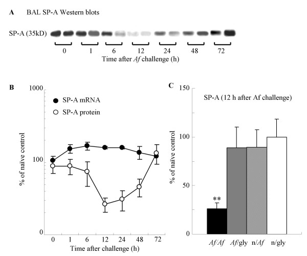
Methods: We studied changes in SP-A expression in the bronchoalveolar lavage (BAL) using a murine model of single Aspergillus fumigatus (Af) challenge of sensitized animals.

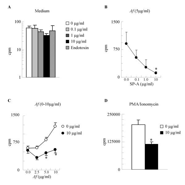
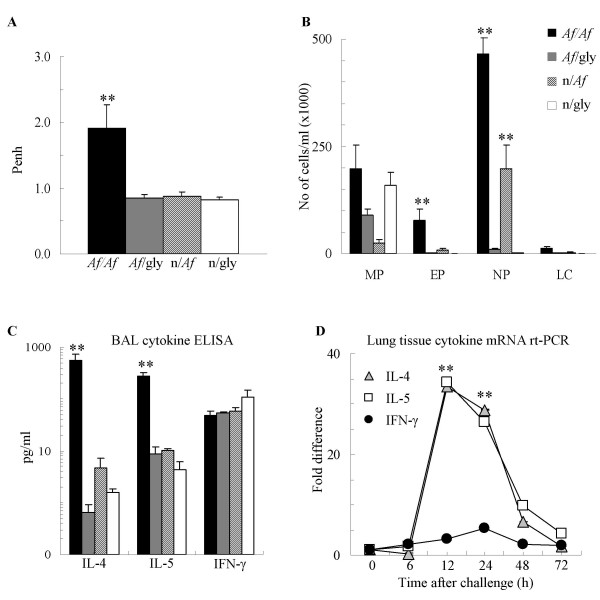
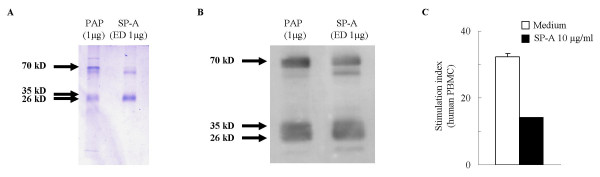
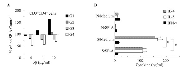
Results: SP-A protein levels in the BAL fluid showed a rapid, transient decline that reached the lowest values (25% of controls) 12 h after intranasal Af provocation of sensitized mice. Decrease of SP-A was associated with influx of inflammatory cells and increase of IL-4 and IL-5 mRNA and protein levels. Since levels of SP-A showed a significant negative correlation with these BAL cytokines (but not with IFN-gamma), we hypothesized that SP-A exerts an inhibitory effect on Th2-type immune responses. To study this hypothesis, we used an in vitro Af-rechallenge model. Af-induced lymphocyte proliferation of cells isolated from sensitized mice was inhibited in a dose-dependent manner by addition of purified human SP-A (0.1-10 microg/ml). Flow cytometric studies on Af-stimulated lymphocytes indicated that the numbers of CD4+ (but not CD8+) T cells were significantly increased in the parental population and decreased in the third and fourth generation in the presence of SP-A. Further, addition of SP-A to the tissue culture inhibited Af-induced IL-4 and IL-5 production suggesting that SP-A directly suppressed allergen-stimulated CD4+ T cell function.

Conclusion: We speculate that a transient lack of this lung collectin following allergen exposure of the airways may significantly contribute to the development of a T-cell dependent allergic immune response.


Figures

Figure 4
Figure 4
SP-A has a significant inhibitory effect on antigen specific lymphocyte proliferation from mice sensitized and challenged with Af. (A): Baseline (unstimulated) proliferation of splenic lymphocytes. Endotoxin in concentration equivalent to the endotoxin content of the 10 μg/ml SP-A samples did not affect cell proliferation. (B): In vitro Af stimulated proliferation of cells from mice sensitized and challenged with Af was inhibited by presence of SP-A, in a dose-dependent manner. (C): The Af dose-response curve of sensitized lymphocytes was abrogated in the presence of SP-A (10 μg/ml). (D) Presence of SP-A inhibited murine lymphocytes stimulated with PMA (2 ng/ml) and ionomycin (100 ng/ml). Background cpm (from wells that contain no cells): 0–16. Mean ± SEM of n = 4 independent experiments presented, each performed in triplicates. * p < 0.05: 0 vs. 10 μg/ml
Figure 1
Figure 1
Allergen challenge of sensitized mice with Af induced a transient decrease in the BAL SP-A protein levels. (A) SP-A Western blots from two representative samples of BAL obtained 1, 6, 12, 24, 48 and 72 h after Af challenge. (B): SP-A protein from BAL (open circles) and lung tissue mRNA (closed circles); % of the naïve control values; n = 4–6. (C): SP-A protein levels measured 12 h after Af challenge Mean ± SEM of n = 8 each; ** p < 0.01 (Af/Af vs. Af/gly).
Figure 2
Figure 2
Single intranasal provocation of sensitized mice with Af induced increase in Penh, inflammatory cell influx and Th2 but not Th1 cytokine release, 12 h After intranasal challenge. (A): Lung function measured by Penh. (B): BAL total and differential cell counts were used to calculate the absolute cell number. MP: macrophages EP: eosinophils, NP: neutrophils, LC: lymphocytes. (C): BAL Cytokine levels measured by ELISA (pg/ml). Mean ± SEM of n = 8 each; ** p < 0.01 (Af/Af vs. Af/gly) (D): Lung tissue cytokine mRNA levels measured by real-time PCR (fold increase in comparison to naïve control levels). ** p < 0.01 (vs. 0 h)
Figure 3
Figure 3
Purification and biological activity of SP-A from human PAP material. (A): Coomassie Blue staining. (B): Western blot comparison of endotoxin-depleted (ED) SP-A preparations (1 μg) with human PAP material (1 μg) used as a positive control. (C): Effect of purified human SP-A on normal human PBMC proliferation. Cells were stimulated with PMA (2 ng/ml) and ionomycin (100 ng/ml). Stimulation Index (cpm of stimulated cells divided by cpm of corresponding unstimulated cells). Mean ± SEM of n = 12
Figure 5
Figure 5
SP-A inhibited Af-induced CD4+ T cell proliferation and release of IL-4 and IL-5. (A): SP-A (10 μg/ml) increased the number of cells in the G1 (black bars) and decreased them in the G3-G4 populations of CD3/CD4+ lymphocytes (% change from the medium-treated samples). Average of 3 independent experiments. (B): Af-induced production of IL-4 and IL-5 by sensitized lymphocytes (S/Medium) was reduced in the presence of 10μg/ml of SP-A (S/SP-A) to levels similar to naïve lymphocytes (N/Medium). Lymphocytes were stimulated by Af (1 μg/ml). IL-4 (grey bars), IL-5 (white bars) and IFN-γ (black bars). * p < 0.05 S/SP-A vs. S/Medium. Mean ± SEM of n = 4 experiments performed in triplicates..

References

    1. Holmskov U, Thiel S, Jensenius JC. Collections and ficolins: humoral lectins of the innate immune defense. Annu Rev Immunol. 2003;21:547–578. doi: 10.1146/annurev.immunol.21.120601.140954. - DOI - PubMed
    1. Gardai SJ, Xiao YQ, Dickinson M, Nick JA, Voelker DR, Greene KE, Henson PM. By binding SIRPalpha or calreticulin/CD91, lung collectins act as dual function surveillance molecules to suppress or enhance inflammation. Cell. 2003;115:13–23. doi: 10.1016/S0092-8674(03)00758-X. - DOI - PubMed
    1. Larche M, Robinson DS, Kay AB. The role of T lymphocytes in the pathogenesis of asthma. J Allergy Clin Immunol. 2003;111:450–63; quiz 464. doi: 10.1067/mai.2003.169. - DOI - PubMed
    1. Haczku A, Atochina EN, Tomer Y, Cao Y, Campbell C, Scanlon ST, Russo SJ, Enhorning G, Beers MF. The late asthmatic response is linked with increased surface tension and reduced surfactant protein B in mice. Am J Physiol Lung Cell Mol Physiol. 2002;283:L755–65. - PubMed
    1. Atochina EN, Beers MF, Tomer Y, Scanlon ST, Russo SJ, Panettieri RAJ, Haczku A. Attenuated allergic airway hyperresponsiveness in C57BL/6 mice is associated with enhanced surfactant protein (SP)-D production following allergic sensitization. Respir Res. 2003;4:15. doi: 10.1186/1465-9921-4-15. - DOI - PMC - PubMed

Publication types

MeSH terms

Substances