. 2005 Sep;11(9):1407-19.
doi: 10.1261/rna.2830905.

Molecular basis for RNA kink-turn recognition by the h15.5K small RNP protein

Affiliations

Molecular basis for RNA kink-turn recognition by the h15.5K small RNP protein

Lara B Weinstein Szewczak et al. RNA. 2005 Sep.

Abstract

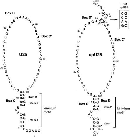
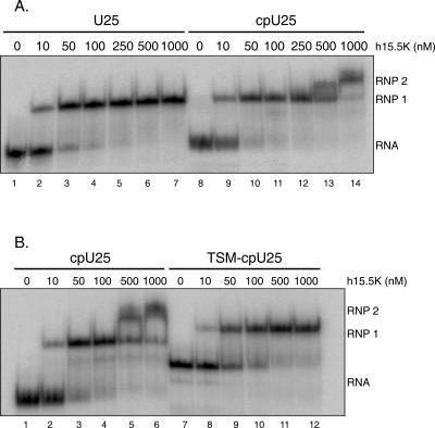
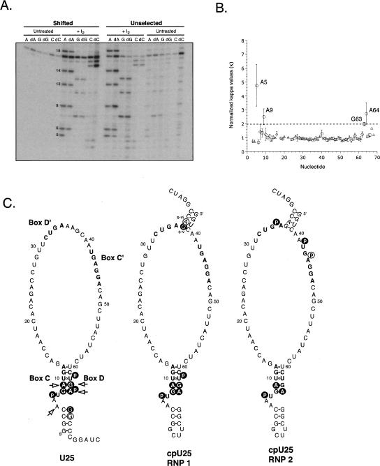
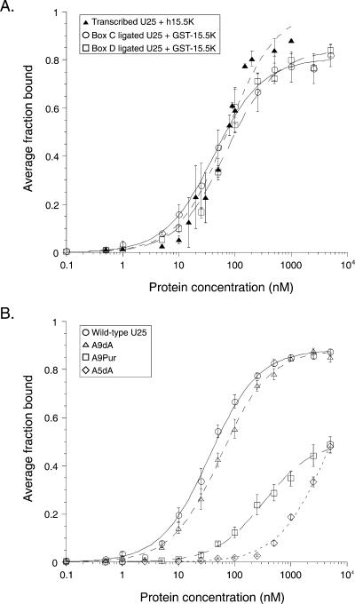
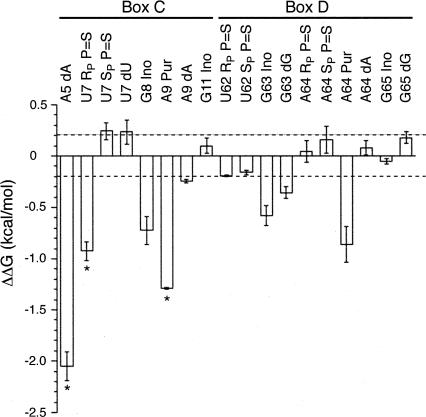
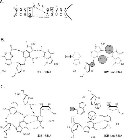
The interaction between box C/D small nucleolar (sno)RNAs and the 15.5K protein nucleates snoRNP assembly. Many eukaryotic snoRNAs contain two potential binding sites for this protein, only one of which appears to be utilized in vivo. The binding site conforms to the consensus for a kink-turn motif. We have investigated the molecular basis for selection of one potential site over the other using in vitro mobility shift assays and nucleotide analog interference mapping of Xenopus U25 snoRNA and of a circularly permuted form. We find that preferential binding of human 15.5K is not dependent on the proximity of RNA ends, but instead appears to require a structural context beyond the kink-turn itself. Direct analysis of the energetic contributions to binding made by 18 functional groups within the kink-turn identified both backbone atoms and base functionalities as key for interaction. An intramolecular RNA-RNA contact via a 2'-hydroxyl may supercede a putative Type I A-minor interaction in stabilizing the RNA-protein complex.


Figures

FIGURE 1.
FIGURE 1.
Secondary structures of U25 and cpU25. Full sequences of Xenopus laevis U25 and cpU25 are shown. Boxes C, D, C′, and D′ are in bold. The 2-nt change disrupting the terminal stem (TS mutant) of cpU25 is shown in the box labeled TSM. CpU25 retains the numbering of U25. Nucleotides added to cpU25 to create the terminal stem are designated s-i through s-xiii, and nucleotides added to seal the U25 termini into a loop are labeled L-i through L-iii. The secondary structure of the box C/D kink-turn motif is shown.
FIGURE 2.
FIGURE 2.
cpU25 forms two complexes with h15.5K. (A) Uniformly [32P]-uridine-labeled U25 (3 nM) forms a single RNP complex when incubated with increasing amounts of h15.5K, while uniformly [32P]- uridine-labeled cpU25 (2.5 nM) shifts sequentially into two complexes, RNP1 and RNP2. (B) Uniformly [32P]-uridine-labeled TSM-cpU25 (7 nM) forms a single complex with h15.5K of different mobility than either RNP1 or RNP2 formed by cpU25 (7 nM).
FIGURE 3.
FIGURE 3.
h15.5K recognizes box C/D in U25, and both boxes C/D and C′/D′ in cpU25. (A) U25 transcripts containing AαS, dAαS, GαS, dGαS, CαS, or dCαS were selected by binding to h15.5K and the shifted RNAs were compared to unselected RNAs after treatment with I2. Substitution of dA at positions 5 and 9 impairs the RNA–protein interaction. dA18 did not cause interference reproducibly. (B) Interference values for dAαS, dGαS, and dCαS are plotted against nucleotide position for U25. Standard errors of the mean are shown for A (circles, n = 4), G (squares, n = 4), C (triangles, n = 2), and U (diamonds, n = 2). (C) Observed sites of interference, which are consistent with h15.5K binding to a kink-turn, are shown for U25 (left) and cpU25 (middle, RNP1 and right, RNP2) as white letters on black circles. Additional sites of interference are circled. Sites where removal of the 2′-hydroxyl caused interference are indicated with arrows. Sequences of the conserved box elements are in bold.
FIGURE 4.
FIGURE 4.
U25 binds equivalently to h15.5K and GST-15.5K. (A) Uniformly [32P]-uridine-labeled U25 binds h15.5K (closed triangles) with an apparent Kd = 73 ± 9 nM in a gel mobility shift assay. Wild-type U25, produced by ligating box C (open circles) or box D (open squares) oligonucleotides, binds GST-15.5K in a filter binding assay with affinities of 42 ± 3 nM or 70 ± 7 nM, respectively. (B) GST-15.5K binds wild type (circles, Kd = 35.9 ± 2.0 nM) and A9dA (triangles, Kd = 57 ± 3 nM) RNAs equivalently in the filter-binding assay. A9Pur (squares, Kd > 330 ± 30 nM) and A5dA (diamonds, Kd > 4200 ± 500 nM) RNAs exhibit substantially weaker binding.
FIGURE 5.
FIGURE 5.
Ribose and phosphate atoms contribute to the U25-GST- 15.5K interaction, assessed by filter binding. The change in free energy of binding relative to wild-type ligated U25 (ΔΔG) is plotted for the individual functional groups measured. Error bars show the standard error of the mean for a minimum of three independent experiments. Asterisks indicate measurements that represent minima. Dashed lines, ΔΔG values > 0.2 kcal·mol−1 or < −0.2 kcal·mol−1 are considered significant.
FIGURE 6.
FIGURE 6.
A tertiary contact promotes K-turn recognition. (A) The primary and secondary structure of the U25 kink-turn is shown. Two proposed tertiary contacts are indicated with dashed lines. (B) (Left) Tertiary contacts between the first bulged nucleotide and the first G-A pair in KT-#7 in the Haloarcula marismortui 50S subunit (Klein et al. 2001). Hydrogen bonds forming both secondary (dashed lines) and tertiary (bold dashed lines) interactions are shown. (Right) Comparable nucleotides from Xenopus U25 are shown. Functional groups with κ values > 2 and contributing > 0.2 kcal/mol to binding are marked with a gray circle outlined in black. (C) (Left) Type I A-minor interaction from the H. marismortui 50S ribosomal subunit (Nissen et al. 2001). (Right) Proposed A-minor interaction in box C/D snoRNAs formed by the second G-A pair and the first base pair in the canonical stem. Functional groups with K values > 2 are in gray circles, as in (A). Functional groups with κ values > 2, but ΔΔG < 0.2 kcal/mol are shown with black circles. Functional groups examined in the NAIM analysis, but showing no interference, are in black boxes in (B) and (C).

References

    1. Batey, R.T., Sagar, M.B., and Doudna, J.A. 2001. Structural and energetic analysis of RNA recognition by a universally conserved protein from the signal recognition particle. J. Mol. Biol. 307: 229–246. - PubMed
    1. Bevilacqua, P.C. and Cech, T.R. 1996. Minor-groove recognition of double-stranded RNA by the double-stranded RNA-binding domain from the RNA-activated protein kinase PKR. Biochemistry 35: 9983–9994. - PubMed
    1. Cahill, N.M., Friend, K., Speckman, W., Li, Z.-H., Terns, R., Terns, M., and Steitz, J.A. 2002. Site-specific crosslinking analyses reveal an asymmetric protein distribution for a box C/D snoRNP. EMBO J. 21: 3816–3828. - PMC - PubMed
    1. Cochrane, J.C. and Strobel, S.A. 2004. Probing RNA structure and function by nucleotide analog interference mapping. Curr. Protocols Nucleic Acid Chem. 6.9: 6.9.1–6.9.21. - PubMed
    1. Cojocaru, V., Nottrott, S., Klement, R., and Jovin, T.M. 2005. The snRNP 15.5K protein folds its cognate K-turn RNA: A combined theoretical and biochemical study. RNA 11: 197–209. - PMC - PubMed

Publication types

LinkOut - more resources